Tuesday, December 1 | 11:50 a.m.-12:00 p.m. | SSG03-09 | Room S404CD
While CT is good at detecting lung nodules, PET/CT is better at determining whether they are malignant or benign. Researchers from Kobe, Japan, wanted to see if their CT technique, known as dual-point contrast-enhanced dual-energy CT (DECT), could rival PET/CT for predicting malignancy."No one has directly compared the capability of dual-energy CT for lung nodule assessment" with PET/CT -- "although it has been suggested as a new technique for contrast-enhanced CT since 2007," said study co-author Dr. Yoshiharu Ohno, PhD, a professor of radiology at Kobe University.
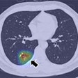
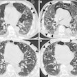
Lead author Dr. Sachiko Miura, Ohno, and colleagues scanned a series of patients with both dual-energy CT and PET/CT, following up with pathological or other exams. With DECT, they generated virtual noncontrast images and iodine maps at both early and late phases, dividing nodules into benign and malignant groups.
Regions of interest placed over the nodules determined the CT values on all images, as well as the differences between early and late phases on virtual noncontrast images. From these, the researchers generated feasible threshold values for positive and negative findings. Meanwhile, maximum standardized uptake values (SUVmax) were also determined for nodules at PET/CT.
DECT was at least as accurate as PET/CT for distinguishing benign from malignant nodules, the group found.
"This study directly compared the diagnostic capability of dual-energy CT for lung nodules with FDG-PET/CT, and demonstrates its utility in routine clinical practice," Ohno said.
