Researchers in Brazil reported the appearance on CT scans of brain anomalies caused by the Zika virus in a letter to the editor published online April 6 in the New England Journal of Medicine.
Only limited imaging data are available about the brain anomalies that may be associated with intrauterine Zika virus infection, noted lead author Dr. Adriano Hazin from the Instituto de Medicina Integral Professor Fernando Figueira in Recife, Brazil, along with several colleagues including Dr. Andrea Poretti from Johns Hopkins University, who provided AuntMinnie.com with the head CT images in this article.
Zika is a mosquito-borne flavivirus that began appearing in Brazil last May. It has been associated with an increasing number of neonates with congenital microcephaly in regions of Brazil where the virus is prevalent, the authors wrote (NEJM, April 6, 2015).
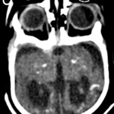
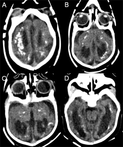
 Axial head CT images of an infant with congenital microcephaly associated with intrauterine Zika virus infection show punctate and larger calcification with a band-like distribution periventricularly, and at the level of the corticomedullary junction within the frontal, parietal, and temporal lobes bilaterally. Ventriculomegaly and global cortical hypogyration are also noted. Image courtesy of Dr. Andrea Poretti.
The group reported what appeared to be the effects of the Zika virus on brain anatomy, as seen in head CT images of 23 infants, obtained at a mean age of 36 days after birth (range, 3 days to 5 months). Intracranial calcifications were seen in all of the infants and mainly involved the frontal lobe (in 69% to 78% of the infants) and the parietal lobe (in 83% to 87%).
The calcifications were mostly punctate (72% to 100%) in configuration, with a predominantly band-like distribution in 56% to 75%. Calcifications were found in the basal ganglia (57% to 65%) and in the thalamus (39% to 43%), the authors wrote.
Ventriculomegaly was seen in all of the infants and was rated as severe in the majority (53%), but it involved only the lateral ventricles in 43%. In addition, all infants had global hypogyration of the cerebral cortex, which was severe in 78%.
Cerebellar hypoplasia was present in 17 cases (74%) and involved only one cerebellar hemisphere in three infants. In two infants, the brainstem was globally hypoplastic. All infants showed abnormal hypodensity of the white matter, and in 87% it diffusely involved all of the cerebral lobes. One infant demonstrated chronic encephalomalacic changes from ischemic stroke in the vascular territory of the left middle cerebral artery.
Intrauterine infection with the Zika virus appears to be associated with severe brain anomalies, including calcifications, cortical hypogyration, ventriculomegaly, and white-matter abnormalities, though it's uncertain when exactly the infection may have occurred in these infants, the authors concluded.
"Our findings are nonspecific and may be seen in other congenital viral infections," they wrote. "The global presence of cortical hypogyration and white-matter hypomyelination or dysmyelination in all the infants and cerebellar hypoplasia in the majority of them suggest that [the Zika virus] is associated with a disruption in brain development rather than destruction of brain. The neuronal and glial proliferation as well as neuronal migration appear to be affected."
The mothers of the infants had symptoms that were compatible with Zika virus infection such as low-grade fever and cutaneous rash during the first or second trimester of pregnancy, similar to findings in other studies, they wrote.


