The use of breast shields during coronary CT angiography (CCTA) studies in women had no effect on the amount of DNA damage patients sustained during exams, but it did degrade image quality, according to a new study in the October issue of Radiology.
Investigators from Walter Reed National Military Medical Center in Bethesda, MD, found similar numbers of DNA double-strand breaks caused by radiation with or without use of the shields in a population of 101 women who had CCTA scans. However, the amount of DNA damage did correlate with the radiation dose level and imaging method used (Radiology, Vol. 281:1, pp. 62-71).
 Dr. Michael Cheezum from Walter Reed.
"At current low levels of radiation, it's important based on these findings to continue to optimize CT protocols, but there is no evidence to support breast shielding," said lead author Dr. Michael Cheezum in an interview with AuntMinnie.com.
Uncertain value
Today's CCTA scans are acquired at far lower doses than in the past, reducing the risk of harmful effects in patients. But not all patients can benefit from low-dose imaging, whether due to high body mass index, arrhythmia, high heart rate, or the use of older CT technology among some providers.
Prior studies using breast shielding showed dose reductions of 30% to 60%, but the data have been limited to phantom models, and there are no studies on the direct biologic effects of breast shielding in women getting scanned. As a result, the use of breast shields is not recommended as a routine tool for reducing radiation dose.
This study aimed to answer the shielding question by measuring the effects of radiation on cellular injury in women undergoing coronary CTA, according to the authors.
Cheezum and colleagues used immunofluorescence imaging of the histone gamma-H2AX, a process that quantifies the number of DNA double-strand breaks with a linear dose response to radiation exposure. They measured double-strand breaks from drawn blood and from an external vial of blood.
Along with the blood draws before and after scans, "we used one of the baseline blood tubes and placed it at the sternum level with and without shielding, so we measured both the amount of in vivo and ex vivo damage resulting from diagnostic imaging," Cheezum said.
Scanned with or without
Women referred for CCTA were randomized to groups in which bismuth breast shields were used (n = 50) or not used (n = 51). The researchers acquired peripheral blood samples before and 30 minutes after in vivo radiation during CT angiography, along with assessing the vials of blood taped to the sternum. They used linear regression and independent sample t-tests to analyze the results.
CCTA scans were acquired on a 64-detector-row scanner (Discovery CT750 HD, GE Healthcare). Most patients (96%) underwent initial noncontrast imaging using prospective electrocardiogram triggering, with a 120-kV tube potential and fixed tube current. Of the 101 patients, 100 had prospectively gated CCTA, and images were reconstructed on a PACS network with a limited field-of-view to obscure any shielding.
The group reconstructed axial CCTA images with a section thickness of 0.625 mm in overlapping 0.4-mm increments using 50% adaptive statistical iterative reconstruction (ASIR, GE). The images were interpreted in consensus by an experienced radiologist and cardiologist. Plaque was graded as normal, no stenosis, nonobstructive, obstructive, or uninterpretable. Radiation dose was measured and the effective dose was calculated.
No effect on DNA damage
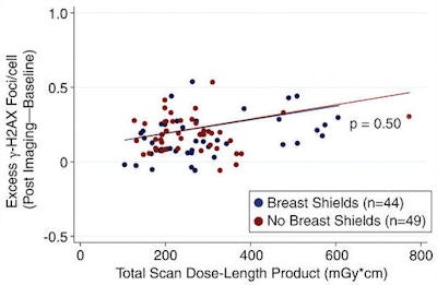
The results showed that breast shielding had no effect on DNA double-strand breaks -- either at the ex vivo breast-tissue level (unadjusted regression: β = 0.08; p = 0.43 versus no shielding) or for circulating blood lymphocytes (β = -0.07; p = 0.50).
"We actually found no evidence of a biologic benefit with breast shielding, and if you compare that to findings in other studies that just optimized CT settings, authors have shown that lowering the radiation dose does lower the amount of gamma-H2AX in circulating lymphocytes," Cheezum said. "The take-home here is that it's really important to lower our dosage in diagnostic imaging, particularly cardiovascular CT, to doses as low as reasonably possible, and to limit the potential for radiation damage."
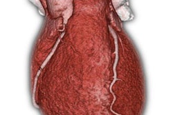
 Images courtesy of Dr. Michael Cheezum.
Among the predictors of increased DNA double-strand breaks were total radiation dose, increasing tube potential, and tube current (p < 0.05). With current radiation exposures (median, 3.4 mSv), breast shielding yielded a 33% increase in image noise and 19% decrease in excellent-quality images, the authors wrote.
Low DNA damage levels overall were likely due to the low radiation doses from prospective gating and dose optimization, Cheezum said, and there were no apparent effects on double-strand breaks from the temperature or timing of the samples, among other factors.
"Other studies used about 30 minutes postimaging, so we took that time point," he said. "Unfortunately, we didn't check at multiple time points following CT to get that peak effect ... so this is a limitation of the study." The study is also limited by the lack of tissue correlation, he added.
Given these results, it would be hard to recommend breast shielding for CCTA in women, but there are anatomic areas where shielding may be helpful without degrading image quality, according to Cheezum.
"Why not shield the gonads in young men? It shouldn't impair image quality, and I don't see any recommendations for it," he said.
A study from Massachusetts General Hospital suggests that "just moving the breasts outside the field-of-view of the scan not only reduces radiation to breast tissue but also improves image quality," he added. That technique appears to be effective, especially in women with large breasts.


