A large analysis of pediatric head CT scans across the U.S. showed surprisingly large variations in radiation dose that increased as children grew older, according to results presented at this week's American College of Radiology (ACR) meeting in Washington, DC.
 Dr. Gelareh Sadigh from Emory University.
In the five-year retrospective study, researchers from Emory University in Atlanta and other U.S. academic centers studied radiation dose indices for single-phase noncontrast head CT scans in patients 18 years and under. The results showed a range of more than 200% between lower- and higher-dose scans, and they suggest that participation in a CT dose index registry or other dose-auditing framework can provide lower overall doses for children in all types of practices.
Doses generally increased with the age of the patient up to 18 years, said radiology resident Dr. Gelareh Sadigh from Emory in an interview. And though more analysis is needed, the results indicate that pediatric hospitals almost certainly provide lower doses than other institutions, she added.
Common for bumps and falls
Head CT exams are one of the most common scans in children, but they're challenging to perform at consistently low radiation doses. Keeping the radiation doses as low as reasonably achievable (ALARA) in practice is critical in the pediatric population because children are more sensitive to radiation and its effects due to their longer expected lifespans versus adults.
The study aimed to examine the national variation in estimated radiation dose for pediatric head CT scans occurring between July 2011 and June 2016. The investigators assessed single-phase noncontrast head CT scans in patients up to age 18 using the American College of Radiology (ACR) Dose Index Registry (DIR). They stratified CT dose index volume (CTDIvol) and dose-length product (DLP) levels by patient age.
In all, the researchers analyzed 268,120 CT scans, including 44% coming from female patients and 59% from children older than 10 years. The median CTDIvol was 35.1 mGy (interquartile range: 24.6-48.2 mGy), representing approximately 200% difference between the 25th and 75th percentiles. CTDIvol increased as age increased. The median DLP was 583.6 mGy-cm (interquartile range: 406.5-791.5 mGy-cm).
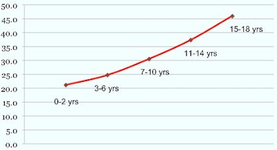 Above, median CTDIvol for head CT by age. Below, median DLP by age. Images courtesy of Dr. Gelareh Sadigh.
There is considerable variation in the radiation dose index for pediatric head CT examinations, the group concluded. These results suggest the benefit of participating in a CT dose index registry, which may provide process improvements in the performance of CT in children across all types of practices.
The head CT doses were almost certainly lower among patients scanned in pediatric hospitals; however, that data cannot be analyzed until the ACR provides information about the origin of each scan, which is expected to be available in the next few months, according to study co-author Dr. Kimberly Applegate, a professor of pediatric radiology at Emory.
"You can expect that when a child goes to a children's hospital, each child is going to get the same thing every time, and that's important," Applegate said, acknowledging that her opinion was based on previous studies, inasmuch as the scan source information was not available for this analysis.
More data analysis needed
Another matter that has not been settled sufficiently is that of children's head sizes. The American Association of Physicists in Medicine (AAPM) has a task group (No. 293) with Image Gently to create size-specific dose estimates (SSDEs) for pediatric head scans, said Applegate, who is a member of the group.
"We have the SSDE measurement for the chest, abdomen, and pelvis, and we're now doing it for the head because it does matter," she said. "It's sort of the last body part to do a size correction for, because the head does change, especially during the first five years of life."
Finally, more information on the individual scanner models is forthcoming in a paper.
"Modern-day scanners are all pretty good" in terms of radiation dose, she said.
A solid start
Considering where we are in dose optimization, the wide variation seen in the study is not too alarming or extreme overall, Applegate said.
"We're all trying to do the best we can, and this gives us good information to work on the next step," she said. "I don't expect my adult [patient] colleagues to work at the pediatric level -- I don't that's a realistic expectation -- but I do expect them to keep an eye on what they're doing."
The point is that everyone who performs CT scans should be in some sort of audit process to know how they're doing, Applegate said. To that end, Image Gently's "Think-A-Head" Campaign? is an important resource for optimizing head CT, she added.
Also, it will be helpful once all of the data are published by institution and the readers from the various institutions are made aware of their results; they can work from there to optimize exposures, Sadigh said.


