An ultralow-dose CT protocol involving computer-aided detection (CAD) matched the accuracy of standard CT for identifying lung nodules at just 10% of the radiation dose of a standard scan, according to an article published online July 25 in the Journal of Thoracic Imaging.
Recent concerns over CT radiation have led to the development of ultralow-dose CT protocols as potential alternatives to conventional CT. Some techniques have dramatically lowered CT radiation dose -- almost to the level of x-rays -- in the evaluation of lung disease.
Researchers from the Mayo Clinic in Rochester, MN, tested a protocol that used lower tube energy and current with tin filtration and a model-based iterative reconstruction protocol (ADMIRE, Siemens Healthineers) to dramatically reduce radiation dose, down to a CT dose index volume (CTDIvol) of 0.4 mGy for a chest scan, compared with 5.3 mGy for a standard scan. To counter image noise and improve sensitivity, they applied CAD (syngo.CT CAD, VA 20, Siemens Healthineers) to the scans.
To assess the combination of the ultralow-dose protocol with CAD for pulmonary nodule detection, they prospectively acquired both conventional CT and ultralow-dose CT scans of 55 patients at their institution between August 2014 and January 2015.
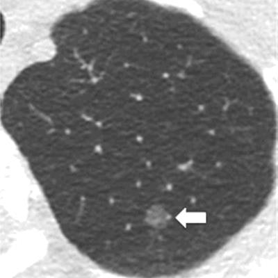
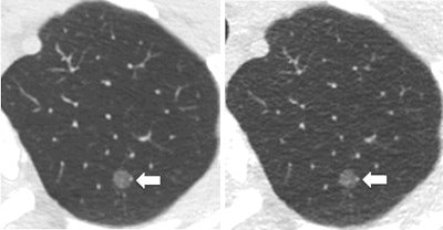
 Standard dose CT (left) and ultralow-dose CT (right) provide comparable visibility of a left upper lobe ground-glass nodule that was missed by readers on the ultralow-dose CT scan but detected by CAD. Images courtesy of Dr. Chi Wan Koo.
Two of four thoracic radiologists evaluated each of the scans; readings by a separate radiologist with more than 20 years of clinical experience served as the reference standard. The radiologists conducted a secondary interpretation of the same scans six months later.
The group found no statistically significant difference between the overall accuracy of the two imaging methods, though there was wide variation in reporting among different readers. The researchers additionally examined the effect of using CAD to perform the same task and discovered that CAD spotted previously undetected nodules about as well as the radiologists did in their second reading.
| Detection of pulmonary nodules on CT and ultralow-dose CT with and without CAD | ||||
| No CAD | CAD | |||
| Standard CT | Ultralow-dose CT | Standard CT | Ultralow-dose CT | |
| Sensitivity | 66% | 60% | 76% | 71% |
The ultralow-dose CT protocol had a comparable sensitivity to standard CT but required a much smaller radiation dose. The mean CTDIvol was 5.3 mGy for standard CT and 0.4 mGy for ultralow-dose CT, and the mean effective dose was 2.8 mSv for standard CT compared with 0.2 mSv for ultralow-dose CT -- a 92.6% reduction in radiation dose (p < 0.0001).
"Ultralow-dose thoracic CT with radiation close to only four chest x-rays' equivalent is achievable without compromising pulmonary nodule detection sensitivity," lead co-author Dr. Chi Wan Koo told AuntMinnie.com. "Computer-aided detection can substantially improve nodule detection sensitivity in these ultralow-dose CT scans for some readers while maintaining interobserver performance."
Increased cancer risk has not yet proved to be true at the low radiation doses associated with medical imaging, noted senior investigator Cynthia McCollough, PhD. "Nonetheless, it is always important to use the lowest doses of radiation that will result in accurate results so that the lifesaving benefits of medical imaging can be used when needed, without fears of accumulating higher doses of radiation," she stated.


