Long before the advent of modern-day imaging modalities, clinicians used manikins to study medical anatomy. Researchers from Duke University will share how they used micro-CT to uncover the internal features and material composition of these human figurines in a presentation at the RSNA 2019 meeting in Chicago.
The earliest accounts of manikin use for medical education date to the late 17th century, when physicians in Germany used the miniature human figurines to study medical anatomy and serve as a teaching aid for pregnancy and childbirth, presenter Dr. Fides Schwartz noted in a statement released by RSNA.
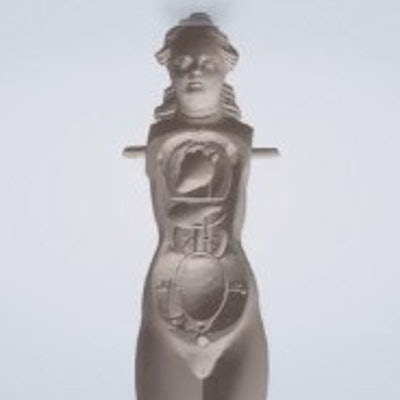
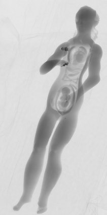 Micro-CT scan of a manikin showing its internal organs and fetus. All images and video courtesy of RSNA.
Researchers have shown interest in studying these manikins. But there are only 180 left in the world, and their fragility and small size have made it difficult to examine them beyond external visualization. Recent advancements in imaging technology, notably micro-CT, have now opened the door for a more detailed look inside the manikins.
For the current study, Schwartz and colleagues used micro-CT to scan a collection of 22 manikins owned by Duke University. They found that most of the manikins contained a variety of internal organs with intricately carved features behind a removable abdominal and chest wall, including the lungs, intestines, and uterus, as well as a fetus visible within the uterus.
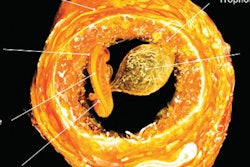
The micro-CT scans also captured volumetric information about the microstructure of the manikins, revealing that 20 of the manikins were made of true ivory, one of ivory and whale bones, and one of antler bones. Several of the manikins contained metallic components and fibers, and as many as 12 manikins contained hinging mechanisms likely held in place by glue or pins.
"The advantage of micro-CT in the evaluation of these manikins enables us to analyze the microstructure of the material used," Schwartz said. "Specifically, it allows us to distinguish between 'true' ivory obtained from elephants or mammoths and 'imitation' ivory, such as deer antler or whale bone."
Looking ahead, the group plans to create virtual 3D models of the manikins in order to produce 3D-printed models for continuing studies.
"This is potentially valuable to scientific, historic, and artistic communities, as it would allow display and further study of these objects while protecting the fragile originals," she said. "Digitizing and 3D printing them will give visitors more access and opportunity to interact with the manikins and may also allow investigators to learn more about their history."


