Artificial intelligence (AI)-based assessment of body composition biomarkers on abdominal CT exams can more accurately stratify risk for future serious adverse events or death in asymptomatic patients than traditional clinical parameters, according to research published online March 2 in Lancet Digital Health.
A team of researchers from the University of Wisconsin School of Medicine and Public Health and U.S. National Institutes of Health (NIH) Clinical Center retrospectively compared the prognostic ability of AI algorithms previously developed by the NIH with that of the Framingham Risk Score and body-mass index (BMI) in over 9,000 individuals receiving abdominal CT exams for routine colorectal cancer screening. They found that the AI algorithms performed better in predicting future serious cardiovascular events and overall survival.
"Given the many millions of CT scans performed each year in many countries, harnessing these valuable data could identify many presymptomatic patients who are at high risk of future serious adverse events, potentially allowing for earlier intervention and prevention," wrote the authors, led by Dr. Perry Pickhardt of the University of Wisconsin School of Medicine and Public Health and senior author Dr. Ronald Summers, PhD, of the NIH Clinical Center.
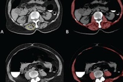
Essentially every abdominal CT scan -- regardless of clinical indication -- contains additional and objectively quantifiable body composition data such as bone mineral density, vascular calcification, muscle mass and density, visceral and subcutaneous fat, and liver fat content, according to the researchers.
"If properly leveraged, these additional opportunistic data could further augment the value of CT scans for the benefit of patients by potentially providing risk stratification for future adverse events and overall mortality," the authors wrote.
The researchers applied five automated algorithms for measuring body composition -- aortic calcification, muscle density, ratio of visceral to subcutaneous fat, liver fat, and bone mineral density -- to the abdominal CT scans of 9,223 people who underwent colorectal cancer screening between April 2004 and December 2016.
All combinations of the AI-acquired CT biomarkers outperformed both the BMI and the Framingham Risk Score for predicting overall survival and major cardiovascular events, as measured by area under the curve (AUC) (p < 0.05).
| Value of AI analysis of body CT scans for predicting overall survival | |||||
| BMI | Framingham Risk Score | AI assessment of aortic calcification and muscle density | AI assessment of aortic calcification, muscle density, and liver density | AI assessment of aortic calcification, muscle density, liver density, and visceral-to-subcutaneous fat ratio | |
| 2-year survival | 0.546 | 0.70 | 0.780 | 0.811 | 0.817 |
| 5-year survival | 0.499 | 0.688 | 0.768 | 0.782 | 0.789 |
The researchers found similar results for the algorithms in predicting future major cardiovascular events.
"This study demonstrates the potential value of harnessing the rich biometric tissue data embedded within all body CT scans that typically go unused in routine practice," the authors wrote. "Although such an opportunistic approach can be applied using manual or semiautomated measures, the maturation of robust, fully automated AI algorithms provides for a more efficient and objective means for high-volume, population-based opportunistic screening."


