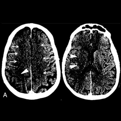
Some of the first CT angiography images documenting the rapid progression of acute ischemic stroke in a patient with COVID-19 were published on May 14 in the American Journal of Neuroradiology (AJNR).
More and more evidence is emerging about the link between COVID-19 and the risk of stroke, particularly among individuals under the age of 50. A definitive cause is still unknown, but patients with severe cases of COVID-19 appear to be at a greater risk for blood clots.
A reason why this may be the case could be that the SARS-CoV-2 virus directly targets the endothelial cells that line blood vessels, which may also put a patient at risk for stroke, according to a study published earlier this month in the Lancet (May 2, 2020, Vol. 395:10234, pp. 1417-1418).
The current case study from Allegheny Health Network Stroke Center in Pittsburgh is of a 64-year-old man who presented to the emergency department after waking up at home with symptoms of left-sided paralysis and shortness of breath. He had tested positive for COVID-19 16 days prior and was recovering well before his sudden onset of ischemic stroke. The patient passed away from complications of COVID-19 three days after admission to the hospital.
 Image A: Noncontrast CT on the day of admission demonstrates subtle findings of acute ischemia in the right middle cerebral artery (arrowheads) and bilateral anterior cerebral artery (arrows) territories, including hypoattenuation and loss of gray-white differentiation. Image B: Repeat noncontrast CT on hospital day two demonstrates progression of acute infarcts in the right middle cerebral artery and bilateral anterior cerebral artery territories, including worsening edema and mass effect. Image courtesy of AJNR.
Data about COVID-19 gathered around the world suggests 5% to 6% of patients with severe cases may suffer a cerebrovascular injury.
"What's unique with our case is that the patient represented a subgroup where a stroke may occur in someone with atypical symptomatic onset," said Dr. Michael Goldberg, a neuroradiologist and director of the Allegheny Health Network Division of Neuroradiology.
The researchers plan to focus their efforts to better treat cerebrovascular injury, they said.


