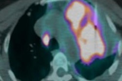
FDG-PET/CT can detect breast cancer metastases predicted by elevated tumor marker values in asymptomatic patients who have already undergone locoregional treatment, according to a study published June 9 in Oncology.
The findings demonstrate how FDG-PET/CT can help clinicians make the clinical information offered by tumor marker data more effective and therefore develop appropriate treatment for breast cancer recurrence, wrote a team led by Dr. Giovanni Corso, PhD, of the European Institute of Oncology in Milan, Italy.
"Since the sensitivity of currently available tumor markers is poor, international guidelines do not recommend their use in clinical practice," the group wrote. "However, clinicians may use them to monitor response, and if they are elevated, imaging staging is often performed. FDG-PET/CT ... is an integrated imaging method useful to detect early locoregional relapse and/or distant metastases."
Same-side breast cancer recurrence after local treatment occurs in about 7% of patients, but the rate of both local and distant recurrence is much higher, at 35%, the researchers noted. Early identification of breast cancer recurrence is key to establishing an effective treatment strategy; women are tracked via clinical examination, imaging, and blood tests that check for biological tumor markers.
Corso and colleagues sought to evaluate how useful blood markers are in predicting distant breast cancer metastases and prompting imaging follow-up with FDG-PET/CT. The group conducted a study that included 561 patients who had surgery for invasive breast cancer and increased tumor marker values on blood testing (antigen CA 15-3 and carcinoembryonic antigen, or CEA) after they completed localized treatment. All of the women underwent FDG-PET/CT in response to the elevated tumor marker data; there were 381 metastases. Of these, 68.5% were bone and/or liver lesions.
The researchers found that increased levels of both tumor markers had strong association with the presence of distant metastases that were confirmed on FDG-PET/CT. The association between CA 15-3 was stronger for bone and/or liver metastases than other types. All results were statistically significant.
| Levels of tumor markers on FDG-PET/CT, with and without breast cancer metastases | ||
| Tumor marker (median value) | No distant metastases | Distant metastases |
| CA 15-3 | 35 U/mL | 58.9 U/mL |
| CEA | 6.6 U/mL | 12.4 U/mL |
The results are in line with previous research that supports the use of FDG-PET/CT for breast cancer patients with elevated tumor markers, Corso et al wrote.
"In these patients, its positive predictive value and accuracy of detection of distant metastases are 97% and 83% to 86%," they wrote. "Moreover, PET/CT can be useful for identifying the site of relapse when traditional imaging is equivocal or conflicting."
Higher tumor marker levels during or after local treatment in asymptomatic breast cancer patients suggest that these women should get additional diagnostic imaging with FDG-PET/CT, the team concluded.
"Specifically, the probability of identifying liver and/or bone metastases is high in cases of increased serum CA 15-3 levels of more than 45 U/ml," the authors wrote.


