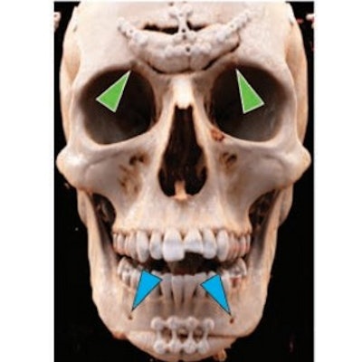
Using CT, radiologists can play a key role in planning facial feminization surgery -- procedures that adjust masculine characteristics such as brow, nose, and chin prominence -- in transgender patients, according to a review published online December 30 in the American Journal of Roentgeonology.
A team led by Dr. Andrew Callen of the University of Colorado Anschutz Medical Campus in Denver offered a list of best practices for preoperative facial feminization surgery planning and stressed how important radiologists are in the care of this patient population.
"Familiarity with [CT] findings will facilitate improved communication between radiologists and surgeons, thereby contributing to the care of transgender women," Callen and colleagues noted.
Many transgender people struggle with dysphoria, or distress due to the mismatch between their birth-assigned sex and the gender with which they identify, the group wrote. Facial feminization surgery is part of a range of gender-affirming surgical procedures. Facial surgery alters characteristics associated with male gender, such as brow prominence and the nasofrontal angle in the upper face, the nasofrontal and nasolabial angles in the middle face, and chin prominence and jaw squareness in the lower face.
Callen's group outlined best practices for radiologists ordering and interpreting preoperative CT exams for facial feminization surgery.
- The CT exam should focus on the complete face and orbits, covering the frontal sinuses through the jaw; acquisition parameters should be a helical thickness of 0.625 mm, kV of 120, and auto mA from 100 to 250. "Maintaining the lowest possible radiation dose ... is of particular importance in the young patient population undergoing facial feminization surgery," the team wrote.
- CT data should be reconstructed using bone kernels in axial, sagittal, and coronal planes.
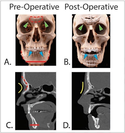
- In the upper face, brow prominence and relationship to the nose are key indicators of male versus female gender identity. "Accordingly, the supraorbital ridge and nose represent major anatomic targets for feminization surgery, and the radiologist has a distinct role in alerting referring clinicians to relevant morphologic findings of these regions," the authors wrote.
- In the midface, the intersection of the frontal and nasal bones and the apex of the nasofrontal angle are important targets of facial feminization surgery. The radiology report should show "presence of a dorsal hump and septal deviation or spurring."
- In the lower face, the goal of facial feminization surgery is to reduce width of the jaw and chin prominence. The lower face radiology report should "describe the location and potential anatomic variations of the inferior alveolar nerve and mental foramina," the group noted.
 Pre- and postoperative CT scans in two different patients undergoing facial feminization surgery. Frontal 3D reformatted images (A) before and (B) after surgery in a 29-year-old transgender woman. Red dashed line indicates frontal bone osteotomy. Red dash-dot lines indicate mandibular angle osteotomies. Red solid line indicates genioplasty. Green arrowheads indicate superior orbital foraminal notches. Blue arrowheads indicate mental foramina. Sagittal reformatted images (C) before and (D) after surgery of 39-year-old transgender woman. Note the Osterhout type 1 frontal sinus. Red dashed line indicates frontal bone recontouring. Red solid line indicates genioplasty. The nasofrontal angle (yellow line) is increased postoperatively. Images and caption courtesy of the American Roentgen Ray Society (ARRS).
Transgender patients often face healthcare challenges, which may include postponed medical care because of a fear of intimidation, harassment, or even refused care, according to Callen and colleagues. Radiologists can help mitigate these challenges.
"[There's] a marked paucity of published transgender-related radiology research, highlighting the need for greater awareness by radiologists of how the specialty may contribute to the care of the transgender community," they concluded.


