Dental surgeons and oral radiologists may be able to detect a serious condition that can cause a stroke as an incidental finding on images taken prior to dental treatment, according to a study published on November 17 in the European Journal of Radiology.
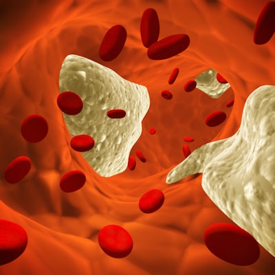
Clinicians may be able to identify atherosclerosis, or plaque buildup, in the carotid artery on conebeam CT (CBCT) scans. This kind of detection can offer dentists the opportunity to aid in the early diagnosis and prevention of strokes, the authors wrote.
"The dental surgeon or the oral radiologist can be the first professional to detect this change and earlier to refer the patient to the physician for further investigations," wrote the group, led by Dr. Heraldo Luís Dias da Silveira, an associate professor of oral radiology at the Faculty of Dental Sciences at Federal University of Rio Grande do Sul in Brazil.
Dental professionals are using CBCT scans more often for various clinical applications in dentistry, including dental implant planning, visualization of abnormal teeth, and endodontic diagnosis. When reviewing these images, examiners will frequently see incidental findings, including periapical, salivary gland, cerebral, and vascular changes.
Among vascular changes, detecting plaque in the internal carotid artery (ICA) as an incidental finding is especially vital for patients. Carotid artery calcification is a known marker of atherosclerosis and is associated with a high rate of morbidity and death. Therefore, the possibility of detecting carotid artery calcification on exams like CBCT that are routinely requested by dental surgeons can affect the general health of patients, according to the authors.
To explore the presence and severity of calcifications on CBCT scans in the extracranial and intracranial pathways of the carotid artery, 284 exams were obtained from a private dental clinic and reviewed by observers who were blinded to any patient characteristics. The patients consisted of men and women who were 40 years and older and who had undergone CBCT scans in preparation for implant placement.
Calcifications were detected in 179 exams (63%), with 57 and 166 in the extracranial and intracranial pathways, respectively. Of the 179 exams, 44 (25%) had calcifications in both pathways, the authors wrote.
Additionally, 19% of calcifications in the intracranial pathway and 60% of those in the extracranial pathway were severe.
Study limitations included the fact that CBCT is not the preferred exam for assessing heart disease. However, CBCT allows detection of calcifications in the extracranial and intracranial pathways of the internal carotid artery, the authors noted.
"There is a high occurrence of ICA calcifications in CBCT as incidental findings in adult patients," the group concluded.


