Wednesday, December 1 | 9:30 a.m.-10:30 a.m. | SSCH06-5 | Room S405
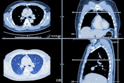
Two ultrashort echo time MRI sequences are comparable to CT when it comes to assessing interstitial lung disease (ILD) in patients with systemic sclerosis, according to this presentation.Presenter Dr. Nicholas Landini of Ca' Foncello General Hospital, Piazzale Ospedale in Treviso, Italy, and colleagues tested two MRI sequences, ultrashort echo time spiral volumetric interpolated breath-hold examination (VIBE) and compressed sensing VIBE, comparing them with standard CT for detecting interstitial lung disease in 54 systemic sclerosis patients and features such as ground-glass opacities, reticulation, honeycombing, and consolidation.
The patients underwent both CT and the two MRI technique exams on the same day. Two radiologist readers assessed the images for ground glass opacities, reticulation, honeycombing, and consolidation; patients were considered positive for interstitial lung disease when ILD extent was more than 5% of total lung volume on visual scores.
Landini's team found that ultrashort echo time spiral VIBE did have some reader variability, but "it could be a reliable tool in assessing ILD and ground-glass opacity extent," with a sensitivity of 95.8% and a specificity of 77.8%, the group wrote. In contrast, compressed sensing VIBE proved an unreliable tool for assessing ILD in patients with systemic sclerosis, with sensitivity of 46.7%.


