Photon-counting CT (PCCT) offers better cardiovascular imaging quality at a similar radiation dose compared with dual-source CT (DSCT) in infants with suspected heart defects, a study published May 23 in Radiology has found.
The findings could improve newborn care, said lead author Dr. Timm Dirrichs of RWTH Aachen University Hospital in Germany in a statement released by the RSNA.
"Infants and neonates with suspected congenital heart defects are a technically challenging group of patients for any imaging method, including CT," he said. "There is a substantial clinical need to improve cardiac CT of this vulnerable group. It's essential to carefully map the individual cardiac anatomy and possible routes of surgical intervention using the highest possible diagnostic standards."
Congenital heart defects are rare -- occurring in only 1% of live births – but they are the leading cause of neonatal morbidity and mortality, the team noted. Of these congenital heart defects, 25% are critical enough to require surgical treatment within the baby's first month. Clinicians assess the defects via ultrasound, MRI, and CT exams to determine whether surgery is needed and/or to create printed 3D reconstructions of the baby's heart.
Dual-source CT technology is typically used for this indication, according to Dirrichs' group, but PCCT offers higher image resolution and reduced radiation dose -- of particular benefit to pediatric patients. Since data on using PCCT for heart imaging in neonates and small children is sparse, the investigators conducted a study that explored use of the technology in this population.
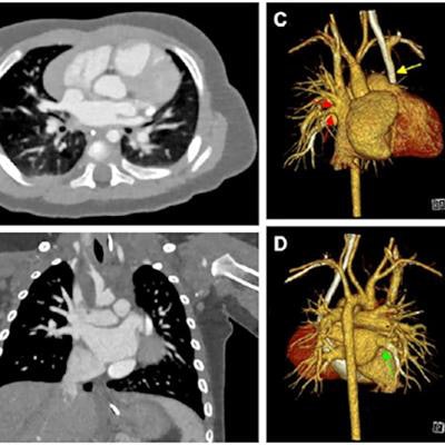
![Cardiac photon-counting CT (PCCT) in a 174-day-old male infant with complex congenital heart defect. (A) Contrast-enhanced axial PCCT image shows sonographically suspected sinus venosus defect with partial anomalous pulmonary venous connection. Soft-tissue window is shown with a 0.6-mm section thickness. Intravenous contrast agent amount: 8 mL of iopromide. (B) Contrast-enhanced coronal PCCT image with soft-tissue window with 0.6-mm section thickness. (C) Anterior view of three-dimensional reconstruction shows a partial anomalous pulmonary venous connection of two separate lung veins from the right upper and middle lobe to the right superior vena cava (red arrows) and a persisting left superior vena cava (yellow arrow). (D) Three-dimensional reconstruction, posterior view. The diagnosis was confirmed at PCCT, and PCCT allowed for visualization of the partial anomalous pulmonary venous connection of two separate lung veins from the right upper and middle lobe to the right superior vena cava. One lung vein drains correctly to the left atrium (green arrow). A sinus venosus defect, an atrial septum defect, an enlarged right atrium, and a persisting left superior vena cava are shown. The only regular confluence of a single right lung vein into the left atrium is shown (D; green arrow). Image quality was rated as 5 of 5 (optimal). Dose-length product was 18. Approximated effective radiation dose, 0.58 mSv (conversion coefficient κ = 0.032 mSv/[mGy × cm]). Images and caption courtesy of the RSNA.](https://img.auntminnie.com/files/base/smg/all/image/2023/05/am.2023_05_23_03_58_3248_2023_05_23_PCCT_infants_figure1.png?auto=format%2Ccompress&fit=max&q=70&w=400) Cardiac photon-counting CT (PCCT) in a 174-day-old male infant with complex congenital heart defect. (A) Contrast-enhanced axial PCCT image shows sonographically suspected sinus venosus defect with partial anomalous pulmonary venous connection. Soft-tissue window is shown with a 0.6-mm section thickness. Intravenous contrast agent amount: 8 mL of iopromide. (B) Contrast-enhanced coronal PCCT image with soft-tissue window with 0.6-mm section thickness. (C) Anterior view of three-dimensional reconstruction shows a partial anomalous pulmonary venous connection of two separate lung veins from the right upper and middle lobe to the right superior vena cava (red arrows) and a persisting left superior vena cava (yellow arrow). (D) Three-dimensional reconstruction, posterior view. The diagnosis was confirmed at PCCT, and PCCT allowed for visualization of the partial anomalous pulmonary venous connection of two separate lung veins from the right upper and middle lobe to the right superior vena cava. One lung vein drains correctly to the left atrium (green arrow). A sinus venosus defect, an atrial septum defect, an enlarged right atrium, and a persisting left superior vena cava are shown. The only regular confluence of a single right lung vein into the left atrium is shown (D; green arrow). Image quality was rated as 5 of 5 (optimal). Dose-length product was 18. Approximated effective radiation dose, 0.58 mSv (conversion coefficient κ = 0.032 mSv/[mGy × cm]). Images and caption courtesy of the RSNA.
Cardiac photon-counting CT (PCCT) in a 174-day-old male infant with complex congenital heart defect. (A) Contrast-enhanced axial PCCT image shows sonographically suspected sinus venosus defect with partial anomalous pulmonary venous connection. Soft-tissue window is shown with a 0.6-mm section thickness. Intravenous contrast agent amount: 8 mL of iopromide. (B) Contrast-enhanced coronal PCCT image with soft-tissue window with 0.6-mm section thickness. (C) Anterior view of three-dimensional reconstruction shows a partial anomalous pulmonary venous connection of two separate lung veins from the right upper and middle lobe to the right superior vena cava (red arrows) and a persisting left superior vena cava (yellow arrow). (D) Three-dimensional reconstruction, posterior view. The diagnosis was confirmed at PCCT, and PCCT allowed for visualization of the partial anomalous pulmonary venous connection of two separate lung veins from the right upper and middle lobe to the right superior vena cava. One lung vein drains correctly to the left atrium (green arrow). A sinus venosus defect, an atrial septum defect, an enlarged right atrium, and a persisting left superior vena cava are shown. The only regular confluence of a single right lung vein into the left atrium is shown (D; green arrow). Image quality was rated as 5 of 5 (optimal). Dose-length product was 18. Approximated effective radiation dose, 0.58 mSv (conversion coefficient κ = 0.032 mSv/[mGy × cm]). Images and caption courtesy of the RSNA.Their study included CT exams of 113 infants who underwent contrast-enhanced PCCT (30), DSCT (83), or both types of exams (one) of the heart and thoracic aorta between January 2019 and October 2022. Median age of the children was 66 days.
The team found that the PCCT images were sharper and showed less noise and more contrast compared with the DSCT images and that readers' mean overall image quality ratings were higher for PCCT than DSCT at similar radiation doses.
| Performance of dual-energy CT compared to photon-counting CT for identifying congenital heart defects in infants | |||
| Measure | DSCT | PCCT | p-value |
| Diagnostic quality image | 77% | 97% | -- |
| Mean overall image quality rating | 3.16 | 4.17 | < 0.001 |
| Signal-to-noise ratio | 29.9 | 46.3 | 0.007 |
| Contrast-to-noise ratio | 37.2 | 62 | 0.007 |
| Mean effective radiation dose | 0.52 mSv | 0.50 mSv | 0.47 |
Any way of improving image quality without boosting radiation dose is beneficial to patients, but especially children, according to Dirrichs.
"PCCT is a promising method that may improve diagnostic image quality and efficiency compared to DSCT imaging," Dirrichs said. "This higher efficiency can be used to reduce the radiation dose at a given image quality level or to improve image quality at a given radiation level."
Further research could explore whether ultrahigh spatial resolution, iodine mapping, tissue characterization, or artifact-reducing algorithms could further improve the image quality of cardiac CT in infants, the authors concluded.


