Understanding the mechanics of flow artifacts on CT or CT angiography (CTA) and how these artifacts are created is key to better disease diagnosis, according to a review published April 25 in RadioGraphics.
In the review, a team led by Caroline Robb, MD, of Mallinckrodt Institute of Radiology, Washington University in St. Louis, MO, described flow artifacts and how they are different from several types of conditions.
"At first glance, flow artifacts may appear as a simple distractor to the discerning eye of a radiologist," Robb and colleagues noted. "However, there are many instances when identification of these artifacts is less straightforward."
Flow artifacts -- that is, "turbulent flow" … "that occurs at the interface of opacified and nonopacified blood" -- are common findings on contrast-enhanced CT and it can be tricky to differentiate from true pathologic conditions, the group wrote.
But recognizing flow artifacts and how they manifest can help radiologists in several ways, including the ability to better identify conditions that predispose patients to these findings, such as pneumonia, chronic lung damage, and altered cardiac output, according to Robb and colleagues.
"By understanding when flow artifacts may be confounding the interpretation of an examination, radiologists can then know when to pursue other troubleshooting methods to assist with the diagnosis," they noted. "In these circumstances, the radiologist can consider several troubleshooting methods, including adjusting the imaging protocols, recommending when additional imaging may be helpful, and suggesting which imaging study would be the most beneficial."
In particular, the group outlined how flow artifacts can be mistaken for pulmonary embolism or vascular injuries:
- Flow artifact versus pulmonary embolism. "When flow artifacts are encountered in the pulmonary arterial tree, they may be mistaken for PE," the team wrote. "There are several characteristics that can be used to distinguish flow artifacts from true PE. True PE will have lower attenuation, with a mean attenuation of 33 HU, while flow artifacts will often have much higher attenuation, with measured values greater than 100 HU."
- Flow artifact versus systemic arterial pathologic conditions. "Flow artifacts can also occur in systemic arteries, most commonly in the aorta," Robb and colleagues noted. "Systemic arterial flow artifacts can cause diagnostic confusion with dissections, vascular injuries, and thrombi. The most common mechanisms for flow artifacts in larger vessels are outrunning the bolus and slow blood flow."
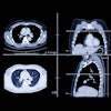
 Flow artifact from slow blood flow along the inner curves of arteries in two patients. (A) Sagittal oblique MPR from a CT examination timed off the aorta in a 78-year-old man with chest pain shows smokelike nonuniform opacification along the inner curve of the descending aorta (arrows), a common location for flow artifacts on CT images. (B, C) Sagittal oblique MPR (B) of the left-sided pulmonary arterial tree in a 76-year-old man with chest pain shows smokelike nonuniform opacification along the inner curve of the posterior basal segment left lower lobe arteries (arrows in B). These findings resolved (arrows in C) on a subsequent CT image (C) with a 70-second delay. Image and caption courtesy of the RSNA.
Yet they can also assist in diagnosis, according to the authors.
"Radiologists can use flow artifacts to localize and characterize vascular defects or determine the direction of blood flow, thereby adding value to imaging examinations," they concluded.
The complete study can be found here.