A deep-learning model applied to CT imaging can predict liver decompensation in patients with primary sclerosing cholangitis (PSC), according to research presented at RSNA 2024.
In a poster study, Yashbir Singh, PhD, from the Mayo Clinic in Rochester, MN, presented his findings, showing that the deep-learning model achieved high marks in predicting liver decompensation. He said these findings highlight the importance of using advanced imaging analysis techniques for better clinical outcomes in patients with chronic liver diseases.
“Clinicians could potentially use this as a tool for early risk stratification, with the model providing predictions approximately 1.5 years before decompensation occurs,” Singh told AuntMinnie.com.
PSC is a chronic liver disease in which the bile ducts inside and outside the liver become inflamed and scarred. Eventually, if left untreated, the bile ducts become narrowed or blocked, leading to cirrhosis.
Singh and colleagues developed and tested a deep-learning model for predicting liver decompensation among PSC patients. The model used CT images to boost early diagnosis and management strategies.
The retrospective study included 277 patients who were diagnosed with large-duct PSC. All patients underwent abdominal CT imaging.
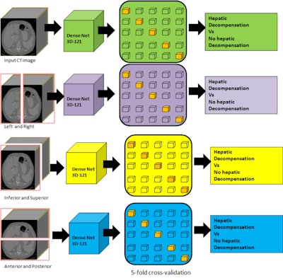
The researchers used portal venous phase images as inputs for a 3D Densenet121 model, trained through five-fold cross-validation. They used several sections of 3D CT images, including the following: right, left, anterior, posterior, inferior, and superior halves. The researchers used this approach to better understand how each anatomic region adds to the model’s predictive accuracy.
The team reported liver decompensation in 128 patients over a median follow-up of 1.5 years.
The deep-learning model scored a baseline area under the receiver operating curve (AUROC) of 0.89. For individual anatomic sections on CT, the model showed varying performance, including the following AUROC scores: left, 0.83; right, 0.83; anterior, 0.82; posterior, 0.79, superior, 0.78; and inferior, 0.76. This suggests that some anatomic regions may be more information for prediction, Singh said.
 Researchers have developed a CT-based deep-learning model for predicting liver decompensation in PSC patients. Pictured is the team's diagram of the pipeline it developed for distinguishing between decompensation and no decompensation is presented in various planes. Image courtesy of Yashbir Singh, PhD.
Singh highlighted that these results show AI’s potential in assisting management of chronic liver diseases by providing early warning of decompensation risk and offering objective, quantitative assessment from routine CT scans. He added that AI could also potentially help stratify patients for clinical trials.
Singh also told AuntMinnie.com that he and his colleagues plan to validate their deep-learning model in a large-scale, multicenter cohort. This includes incorporating topological data analysis and studying whether the model detects PSC-specific features or general cirrhosis features.
He added that future research aims to explore applications for detecting other PSC complications like cholangiocarcinoma and expand this deep-learning approach to other liver diseases like non-alcoholic fatty liver disease.


