Using precontrast low attenuation at CT as an ancillary feature (AF) increases the proportion of liver cancers categorized as LI-RADS-4 and improves the framework's sensitivity for diagnosing the disease, researchers have reported.
"Current CT AFs have limitations, as several … are more evident at MRI," wrote a team led by Rohee Park, MD, of Asan Medical Center in Seoul, South Korea. "Precontrast low attenuation may serve as a potential AF in the Liver Imaging Reporting and Data System (LI-RADS)."
AFs that suggest malignancy on CT may include iron sparing in a solid mass, mosaic architecture, or corona enhancement. Precontrast low attenuation on CT shows up as a region darker than surrounding tissues because fewer x-rays are being blocked, the team explained, and can help identify liver cancer and differentiate it from other conditions.
Park's group evaluated the diagnostic value of precontrast low attenuation as an additional AF for diagnosing liver cancer and its impact on LI-RADS diagnostic performance. The team conducted a study that included 194 adults at risk of the disease who underwent multiphase dynamic liver CT before liver resection or transplant between January and December 2022. From the 194 patients, there were 328 "hepatic observations": 187 (57%) liver cancers, 26 (7.9%) nonliver malignancies, and 115 (35.1%) benign lesions.
Two radiologists evaluated any major and/or ancillary features for each of these found lesions using the following LI-RADS categories: LR-3 (intermediate probability of malignancy), LR-4 (probably liver cancer), and LR-5 (definitely liver cancer). The readers assigned each lesion a LI-RADS category twice, once using AFs only and second using AFs with precontrast low attenuation.
The group reported the following:
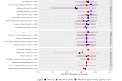
- Precontrast low attenuation was associated with liver malignancies, yielding a diagnostic odds ratio of 9.1 (p < 0.001).
- Adding the precontrast low attenuation technique upgraded 20 lesions (17 cancers and 3 dysplastic nodules) from LR-3 to LR-4, increasing the proportion of malignancies in LR-4 from 64.7% to 72.2%.
It also found that using the precontrast low attenuation CT technique as an ancillary feature improved sensitivity for identifying liver cancer compared with not including it. There was no difference in specificity.
Performance comparison for identifying liver cancer using precontrast low attenuation CT technique as an ancillary feature | |||
Measure | LI-RADS with AFs only | LI-RADS with AFs and precontrast low attenuation | p-value |
| Sensitivity | 79.9% | 88.6% | p < 0.001 |
| Specificity | 85% | 82.7% | p = 0.06 |
"Precontrast low attenuation at CT may be considered to be an additional AF, potentially aiding in the identification of more high-risk lesions and supporting their categorization into LR-4," the researchers wrote.
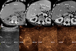
 Axial CT images in a 67-year-old man with hepatitis B virus infection–related liver cirrhosis show a 1.8-cm hepatic observation in segment VIII (arrows). The lesion shows (A) nonrim arterial phase hyperenhancement without nonperipheral washout and it remains (B) mildly hyperenhancing in the portal venous phase and shows (C) isoattenuation in the delayed phase. No ancillary features are visible, and the lesion is categorized as LR-3 according to Liver Imaging Reporting and Data System, or LI-RADS, version 2018. (D) On the precontrast image, the lesion appeared as a low attenuating lesion and was upgraded to LR-4 based on precontrast low attenuation as an ancillary feature favoring malignancy. Pathologic examination after hepatic resection served to confirm hepatocellular carcinoma. Images and caption courtesy of the RSNA.
"Future studies including patients with various liver diseases who have undergone treatments such as ablation or transarterial chemoembolization are needed to more fully elucidate the diagnostic value of precontrast low attenuation at CT and its role in guiding clinical decisions," they concluded.
The full article can be accessed here.