Brazilian researchers have developed a software application that analyzes digital x-ray images to improve the assessment of scoliosis. The researchers believe the software could provide a more accurate method than manual measurements for calculating angles caused by the curvature of the spine.
The severity of scoliosis has traditionally been calculated using the Cobb angle, a measurement of spine curvature on an anteroposterior radiograph. The degree of the Cobb angle can guide patient management, with angles greater than 20° requiring braces and those greater than 40° frequently leading to surgery.
Recently, however, many clinicians have begun using vertebral axial rotation in place of the Cobb angle for assessing scoliosis, according to a paper published online October 10 in the European Spine Journal. Orthopedic templates are overlaid on spine images, and vertebral axial rotation angles are calculated manually to diagnose scoliosis, according to a research team led by Alan Pinheiro and colleagues from the University of São Paulo in São Carlos.
The rise of digital x-ray creates the opportunity that vertebral axial rotation can be calculated automatically via software, however, Pinheiro and colleagues said. They decided to analyze the accuracy of an application that Pinheiro developed by comparing it to the traditional manual method.
The software is based on the methods developed by Perdriolle and Raimondi for measuring axial vertebral rotation. The researchers tested the application in two human vertebrae (T3 and L4) in a lab experiment using a device that rotated the vertebrae around a fixed shaft. Vertebrae were rotated in 13 stages, from 0° to 60° in 5° increments.
Images of the vertebrae were acquired with a conventional analog radiography system. Three orthopedic surgeons analyzed the films and calculated vertebral axial rotation angles using a Pedriolle torsiometer, a Raimondi table, and a ruler. No marks were made on the images.
To enable the images to be analyzed with the software, they were digitized at a resolution of 1,000 x 450 pixels at 300 dots per inch with a commercially available digitizer (Diagnostic Pro, Vidar Systems, Herndon, VA), with 13 acquisitions acquired of each vertebra for a total of 26 images.
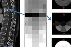
With Pinheiro's software, users mark the pedicle position with a single mouse click, while the width of the vertebra is determined at the narrowest part of the vertebra using a straight line drawn by the observer on the computer screen. This information is sufficient for the application to provide three key analyses:
- Automatic projection of the pedicle distances to the convex vertebral side
- Estimation of actual vertebra width
- Rotation corresponding to the relation between the first two measurements according to the values defined by the Pedriolle and Raimondi methods
The three human observers made a total of 468 measurements of the plain films, matched by an equal number of measurements for the software. The degree of accuracy for both techniques was analyzed using root mean square (RMS) error, while measurement precision was analyzed via standard deviation (SD). Accuracy and precision were calculated for both Raimondi and Pedriolle methods, with the software proving to be more accurate than manual techniques.
Degree of error, manual versus software by measurement method
|
According to Pinheiro and colleagues, the findings indicate that using software to analyze digital images produced angle measurement errors that were 43% smaller than manual errors. They also found that the automated technique had higher correlation coefficients and confidence intervals, indicating more consistent data.
A measurement error of 5° or greater could lead to patient misdiagnosis using the Cobb angle method but, fortunately, is not as consequential using the axial rotation method because three to four vertebra are evaluated at once, helping compensate for random errors, Pinheiro said.
The authors said that there were other advantages to digital analysis of radiography images, including the ability to immediately compare radiological images and easier image storage and transmission. The software also requires little individual intervention, which may help less experienced observers estimate vertebral rotation.
Pinheiro told AuntMinnie.com that the software includes other features, such as the ability to measure Cobb angle, calibrate medical images, and measure angles and distances between points. Although the current study used digitized x-ray films, he believes it could also be used with images acquired with digital radiography (DR) systems.
The software is currently being used at Pinheiro's facility to evaluate patients with scoliosis. The group hopes to validate the application's ability to measure axial rotation, Cobb angle, and other features. Pinheiro said the software could be commercialized at some point in the future.
By Brian Casey
AuntMinnie.com staff writer
October 28, 2008
Related Reading
Surgery for degenerative lumbar scoliosis often effective: review, August 24, 2009
Hard-copy prints help orthopods adjust to digital imaging, August 3, 2009
Study finds lower dose for slot-scanning DR unit, November 3, 2008
Copyright © 2009 AuntMinnie.com
