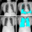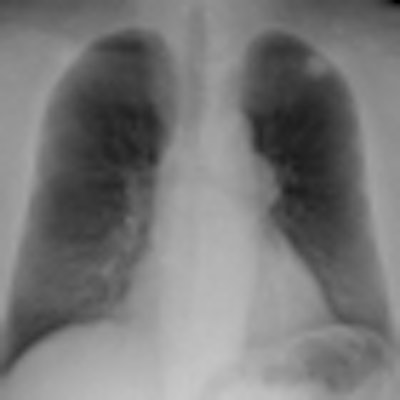
Dual-energy imaging is a promising technique for improving the performance of digital radiography (DR) in finding lung lesions by removing bony obstructions, but it requires specialized hardware. University of Chicago researchers may have found a software-based alternative.
Calling their technique virtual dual-energy DR (or bone suppression imaging), the technology substitutes sophisticated software algorithms for the hardware required to take two projection x-ray studies, match them, and separate out bone that might obscure pathology. Results with the software-based technique showed that it improves upon temporal subtraction, a DR method for tracking changes on radiographs over time, according to the University of Chicago's Kenji Suzuki, Ph.D., who presented results at the 2009 RSNA meeting.
In a related study, Dr. Heber MacMahon, director of thoracic radiology at the University of Chicago, followed Suzuki's presentation with findings indicating that bone suppression imaging improves upon DR alone for detecting small lung cancers, though it remains second best to hardware-based dual-energy imaging for accurately identifying small pulmonary nodules.
Bony obstructions
Dual-energy imaging is being pursued to solve the problem that bony structures pose for projection radiography, where small lung cancers can hide behind a rib or clavicle to mislead a radiologist. MacMahon and colleagues at the University of Chicago have been working on solutions since the installation of their first dual-energy system in 1996. A recent study by the group found that bony structures were partly responsible for eight of every 10 missed lung cancers, Suzuki said. Undetected lung cancer on DR is a major source of malpractice claims against radiologists.
Dual-energy imaging separates the high- and low-energy components of the x-ray beam to produce images of bone without soft tissue, and images of soft tissue without bone. A standard frontal chest x-ray is also produced to characterize both bone and soft tissue, MacMahon said in an interview.
Despite published studies establishing dual-energy imaging's ability to detect small, subtle pulmonary abnormalities, the technique has not been widely adopted, possibly because of the $20,000 to $25,000 extra cost for the specialized hardware or software and the lack of reimbursement for generating and reading two additional images, MacMahon said.
Dual-energy imaging may also involve slightly more radiation than conventional radiographs, especially when used with the conventional two-shot technique that requires two exposures in rapid succession.
To reduce bony artifacts, Suzuki developed a data filter called a massive-training artificial neural network (MTANN). The MTANN was trained on chest radiographs and corresponding soft-tissue images acquired with dual-energy radiography, Suzuki said. After training, the MTANN could generate subtracted soft tissue, bone, and composite chest images without specialized dual-energy hardware.
The University of Chicago has licensed a concept similar to MTANN to Riverain Medical of Miamisburg, OH. After modifications, the firm introduced the product, SoftView, as a work-in-progress at the 2009 RSNA meeting. It is approved for sale in Europe and Canada, while 510(k) clearance from the U.S. Food and Drug Administration (FDA) in the U.S. is pending.
Less artifact, more confidence
In Suzuki's study, the University of Chicago team wanted to see if MTANN could improve upon temporal subtraction, an advanced DR method of tracking interval change in chest radiographs as a sign of possible pathology. The temporal subtraction approach digitally separated bone from soft tissue, but artifacts from bone could still appear because of nagging image registration problems.
Suzuki and colleagues found that a chest radiologist consistently found less residual artifact and expressed more confidence in the clinical utility of virtual dual-energy bone suppression images, compared to conventional temporal subtraction radiographs, in 100 sequential pairs of images acquired from 53 lung cancer patients.
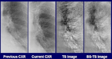 |
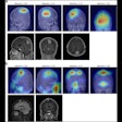
| Radiologists and residents in the study by Suzuki and colleagues rated anatomic changes associated with lung cancer in the bone suppression-temporal subtraction (BS-TS) image as more evident than a temporal subtraction (TS) image derived from previous and current chest radiographs. Image courtesy of Kenji Suzuki, Ph.D., and Dr. Heber MacMahon. |
Images were graded on a five-point scale. In terms of image registration, conventional temporal subtraction was rated at 3.55, compared with 3.91 for virtual dual-energy bone suppression. On the measure of diagnostic confidence, the ratings for conventional temporal subtraction and virtual dual-energy suppression were 3.54 and 3.66, respectively. The differences for both measures were statistically significant.
"The radiologist's evaluation suggests that the contrast of ribs in the original digital chest radiography was reduced substantially, while soft-tissue contrast was maintained," Suzuki said in an interview. "Rib artifact was reduced substantially."
Value of bone suppression
In the other RSNA meeting presentation, a research team led by MacMahon asked three experienced radiologists and two radiology residents to rate their diagnostic confidence in their interpretation of conventional digital chest radiographs, DR performed with Riverain's investigational bone suppression software, and dual-energy imaging with dedicated hardware for detecting nodules. The image set comprised 50 patients harboring CT-confirmed nodules known to be less than 20 mm in diameter, intermingled with studies from 30 healthy subjects without lung cancer.
The findings established that the readers were significantly more confident in their interpretation of the bone suppression images than with standard digital chest x-rays. But they also reported significantly more confidence in their readings of dual-energy imaging than with bone suppression imaging.
 |
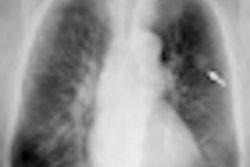
| In the MacMahon et al study, radiologists rated a small pulmonary nodule (< 20 mm) as more conspicuous on a bone suppression image (left) than on standard chest radiograph (center), where it was partly obscured by overlying bones. Dual-energy subtraction (right) provided the most complete bone subtraction but required special equipment to perform. Images courtesy of Dr. Heber MacMahon. |
A receiver operator characteristics (ROC) statistical analysis found that reading accuracy followed the same pattern. Bone suppression via software led to more accurate diagnoses than standard x-rays, but hardware-based dual-energy imaging was even more accurate.
"We are saying that if someone uses dual-energy, it seems to perform better than bone suppression software alone," MacMahon said. "On the other hand, bone suppression provides a software-only solution, and can provide many of the benefits of dual-energy without special equipment."
Findings from both studies were limited by the small size of their patient populations, the high prevalence of lung cancer and other possible selection biases among cases used for interpretation, and data collection from a single imaging service.
University of Chicago researchers associated with the scientific inquiries have received research grants from Riverain Medical and other imaging-related software and device manufacturers. Suzuki received a research grant from Riverain, and MacMahon is a consultant for the company.
By James Brice
AuntMinnie.com contributing writer
February 10, 2010
Related Reading
Rads prefer dual-energy x-ray over standard DR in lung, January 29, 2009
Adding CsI-based dual-energy imaging doesn't improve lung nodule detection, December 26, 2008
Dual-energy digital x-ray still looking for acceptance, February 22, 2007
Copyright © 2010 AuntMinnie.com
