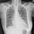
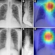
Pairing low-dose digital radiography (DR) tomosynthesis with conventional radiography eliminates the need for three of every four CT scans typically performed to confirm lung lesions previously suspected on chest radiography, according to a study from the University of Trieste in Italy.
Emilio Quaia, MD, an associate professor of radiology, and colleagues at the university's Cattinara Hospital found that, in addition to reducing CT use, performing tomosynthesis after identifying suspicious pulmonary abnormalities with conventional DR exposes patients to less ionizing radiation and shortens the waiting time for follow-up imaging when CT is required. Quaia elaborated on this group's investigations at the 2010 RSNA meeting in Chicago.
The group's findings on the diagnostic power of chest tomosynthesis were first published in the journal Academic Radiology in October (Vol. 17:10, pp. 1267-1274). Based on clinical experience with 228 consecutive patients, the researchers found that tomosynthesis following digital radiography was 90% to 92% accurate among two experienced readers for assessing the possible pulmonary nature of suspicious lung masses.
Only 14 or 251 suspicious pulmonary lesions were miscategorized, with the sources of those errors stemming from an inaccurate appraisal of their location, Quaia said in an interview with AuntMinnie.com. Twelve were pleural plaques, and two were cardiac calcifications. All appeared in the anterior region of the lung parenchyma near the thoracic wall.
"This does not mean that those lesions were missed or not identified by digital tomosynthesis, but the location described by the reader was wrong," Quaia told AuntMinnie.com. "If a pulmonary lesion is misclassified as a pleural lesion, this could potentially lead to underestimation of lung cancer."
The study summarized by Quaia at the RSNA meeting considered the group's experience with 287 consecutive patients. Digital chest radiography was performed on a Definium 8000 (GE Healthcare, Chalfont St. Giles, U.K.). The same system equipped with optional VolumeRAD hardware was used for digital tomosynthesis.
Tomosynthesis resolved doubtful radiographic findings for 75% of the patients (213/287), reducing the need for CT to only 25% (74/287). CT was deemed unnecessary when tomosynthesis clearly showed lesions were benign from the characterization of calcified solid nodules, definite or probable extra-pulmonary nodules, and pseudolesions, Quaia said.
Digital tomosynthesis had a dramatic effect on CT utilization. More than 250 chest CTs were performed on 70 patients with suspicious pulmonary masses in the year before the adoption of digital tomosynthesis in January 2008. During the year after adoption, 85 patients received fewer than 50 CT exams. And fewer than 50 CT studies were performed on 132 patients with suspicious lung lesions examined from January 2009 to September 2010.
Lower CT utilization translated into less radiation exposure for patients who avoided diagnosis with CT. A CT exam involved doses of 1 mSv to 8 mSv depending on the patient's body mass index and the type of scanner used in the procedure. Patients were exposed to a mean dose of 0.2 mSv from digital tomosynthesis and 0.06 mSv from two-view conventional DR, Quaia said.
A shorter waiting time for confirmatory imaging was another advantage of tomosynthesis. Patients underwent tomosynthesis an average of four days (± 1 day) after positive radiography. The average waiting time between tomosynthesis and CT for patients requiring the additional procedure was 15.2 days. Before implementation, CT waiting times averaged 24.6 days after the initial chest x-rays.
For radiologists, tomosynthesis studies took longer to read than conventional DR, but less time than chest CT. The mean reading time for tomosynthesis was 200 seconds (± 40 seconds) compared to 120 seconds (± 30 seconds) for a digital chest radiograph and 10 minutes (± 5 minute) for chest CT, Quaia said.
The study presented at the RSNA meeting confirmed continuing problems using tomosynthesis to characterize pleural lesions, especially in the anterior region of the chest. Reader uncertainty about hilar lesions often led to follow-up CT, Quaia noted.
The limitation most likely stems from the limited 35° x-ray tube scanning range of tomosynthesis compared with the complete 360° x-ray tube and detector rotations during CT. The group is planning to conduct more research to define the variable diagnostic power of chest tomosynthesis by anatomic region, Quaia said.
By James Brice
AuntMinnie.com contributing writer
December 23, 2010
Related Reading
Study finds clinical niche for DR tomo in pulmonary lesions
Digital chest tomo beats standard DR for pulmonary nodules
Will new DR technologies offer alternative to CT? Part 2
Copyright © 2010 AuntMinnie.com
