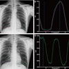
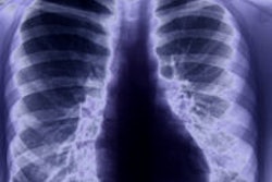
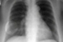
A new study is helping public health officials gain confidence in chest computed radiography (CR) as an alternative to film-screen x-rays for monitoring black lung disease and other forms of pneumoconiosis.
In research published in the December issue of Chest, A. Scott Laney, PhD, and colleagues with the Division of Respiratory Disease Studies at the National Institute for Occupational Safety and Health (NIOSH) determined that CR is at least equivalent to film-screen radiography for recognizing small pulmonary opacities from pneumoconiosis. They also concluded that the image quality of CR is actually better than film-screen radiography (Chest, December 2011, Vol. 140:6, pp. 1574-1580).
These findings are probably not surprising to radiologists who are familiar with digital imaging, but CR performance is still an open question to epidemiologists who manage long-term longitudinal studies of underground miner respiratory health for NIOSH and the United Nation's International Labor Organization (ILO). The ILO classification system has relied on film-screen chest radiography for 80 years.
The system's current application indicates the incidence of black lung disease is rising to epidemic levels, Laney told AuntMinnie.com in an interview. Autopsies on the 38 men killed in West Virginia's Upper Big Branch Mine disaster of 2010, for example, found evidence of pneumoconiosis in 70% of the victims, he said.
Considering the serious public health ramifications, researchers cannot afford to jeopardize the integrity of the international program by converting to an imaging modality that could inadvertently recalibrate the standardized scale for gauging disease incidence and severity.
The need to assess the application and equivalency of the ILO system in the context of digital imaging has become apparent as it has become more widespread, said Alfred Franzblau, MD, the associate dean for research at the University of Michigan School of Public Health.
ILO published revised guidelines in November based on digitized versions of plain film images permitting the application of digital imaging to its scale for diagnosing and characterizing pneumoconiosis. This action is still considered a preliminary step toward the organization's full acceptance of digital imaging, however.
In Laney's trial, storage phosphor CR and standard film-screen chest images were drawn from 172 of 1,401 male underground miners who participated in a previous study derived from NIOSH's Enhanced Coal Workers' Health Surveillance Program.
Both imaging exams were performed the same day and were read twice by so-called B readers, who are certified by NIOSH as competent to evaluate radiological studies of pneumoconiosis. Images were included when one or more original classifications indicated the presence of small opacity profusion.
Findings were based on 2,408 readings (1,204 for each modality). Image quality was judged on a four-point scale from "good" to "unacceptable."
Using these methods, Laney and colleagues identified little meaningful difference between the two modalities for visualizing small pneumoconiotic opacities.
CR produced higher quality images than film-screen radiography. Researchers found that 93.4% of the CR studies were classified as either "good" or "acceptable, no defects," compared with 79.6% of the film-screen studies.
Film-screen imaging uncovered more small opacities (profusion category ≥ 0/1) than CR, but intermodality agreement was deemed moderate to good relating to profusion category 0/0 classifications from an initial and repeat reading of the images.
Significantly more irregular opacities, such as retricular structures, were characterized with CR, compared with the typically rounded features of opacities that appeared on the film-screen images. CR was also more likely to detect opacities with diameters smaller than 1.5 mm.
Laney, who has been involved in several similar trials, believes the current study offers a definitive answer about equivalence between the two modalities.
"This puts the icing on the cake saying that we really can move to adopt digital technologies," he said.
Franzblau noted the results build on and extend prior work in this area. The study design and statistical methods are appropriate, and the conclusions are well-supported by the data and analyses, he said.
"This study increases our confidence in the use of the ILO system as we continue to move ahead with use of various forms of digital radiography in the realm of pneumoconiosis," Franzblau said.


