New forms of image artifacts can compromise the performance of digital radiography (DR) equipment, say researchers from the Mayo Clinic in Rochester, MN, in the January issue of the American Journal of Roentgenology. The group described its experience with DR artifacts and how to correct for them.
Imaging facilities are shifting to DR equipment due to its inherent workflow efficiencies compared to analog x-ray, or even computed radiography (CR). But DR's recent growth means that many facilities are encountering artifacts that are novel to the technology.
"These are things users need to be aware of, especially things that could jeopardize patient safety," said Alisa Walz-Flannigan, PhD, lead researcher on the study (AJR, January 2012, Vol. 198:1, pp. 156-161).
Artifacts featured in the Mayo paper were produced by flat-panel DR detectors with an amorphous silicon thin-film transistor array, coupled to a phosphor layer of either cesium iodide or gadolinium oxysulfide. Specific equipment models in the study included DRX-1 from Carestream Health, Definium 8000 from GE Healthcare, DigitalDiagnost from Philips Healthcare, and Axiom Aristos MX from Siemens Healthcare.
Similar problems have not affected Mayo's CR systems because of essential differences in detector design between the CR and DR systems. Mayo's experience varied with the different makes and models of DR equipment.
In an interview with AuntMinnie.com, Walz-Flannigan discussed the implications of image lag artifact, a problem linked to DR detector design, and backscatter artifact, an issue primarily associated with inadequate radiation shielding for wireless digital detectors.
Artifacts due to image lag (also referred to as ghosting) have raised safety concerns. Walz-Flannigan pointed to instances where the shadows of lead laterality markers positioned on a patient during an earlier procedure reappear as artifacts on subsequent images.
"This could have led to wrong-side labeling," she said.
Ghosting seems to arise from an inability to deactivate detector plate phosphors that were energized on a completed image before later image acquisitions are performed. The trapped charges produce a double exposure, making the next image in a series either difficult or impossible to read.
 |
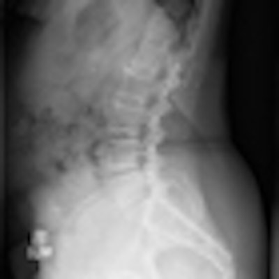
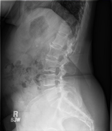
| Outline of the patient's neckline and lead marker from a swimmer’s view of a shoulder study are seen (with inverse opacity) in this lumbar spine image taken two minutes later. The most visible artifact is directly posterior to the lumbar spine. All images courtesy of Alisa Walz-Flannigan, PhD. |
Highly opaque objects depicted in a DR image will reappear less densely in one or more subsequent images. Lag artifacts typically do not appear in images acquired 60 seconds after their onset, but intense opacities did persist sporadically for up to 15 minutes after image acquisition on a particular DR unit at Mayo because of a manufacturer's unsuccessful attempt to use a subtraction technique to eliminate the phenomenon.
"This is when we realized the vendors were actually doing something actively," Walz-Flannigan said. "Otherwise, we would have never gotten this strange artifact."
Working with the vendor, Mayo staff devised strategies that have helped to mitigate the artifact problem -- but at a cost of reduced machine efficiency.
The order in which images are acquired was modified to avoid intense activation of the digital plate quickly followed by another acquisition. Technologists are instructed to pause for at least a minute between high-exposure/high-contrast scans for detector recovery, and they are encouraged to consult with radiologists to determine whether to salvage or retake damaged views.
"Any efficiency gain we got from DR was eaten away by the fact that we now have to pause after some types of exposures," Walz-Flannigan said.
DR vendors have pursued software solutions to find proprietary ways to eliminate lag artifacts. Some revisions have worked so well that lag is no longer measurable, she noted. And vendors have been instrumental in improvising fixes for severe backscatter artifacts that have appeared on images acquired with Mayo's wireless digital detectors.
 |
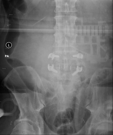
| Backscatter artifact expressed itself as an outline of DR system electronics imposed on high-exposure images of large patients or long exposures. |
Mayo physicists fitted a phantom with pads of synthetic fat to reproduce the artifact under controlled conditions.
Based on this inquiry, they discovered that a lack of lead shielding -- possibly removed from the detector to reduce its weight and make it more marketable -- produced conditions that allowed the backscatter artifact to appear.
The problem disappeared when a lead apron was draped over the detector. The vendor then responded by taping a thin sheet of lead to the back of the detector.
Walz-Flannigan now considers the backscatter problem solved.
"The vendors just need to be cognizant that you can only reduce the detector's weight so much," she said. "Otherwise, the system is susceptible to artifacts."
On the clinical side, radiologic technologists, in general, need to be informed about the unique conditions that apply to DR operation, she said. Those issues will be addressed in new DR quality control guidelines that will soon be published by the American Association of Physicists in Medicine.


