South Korean researchers have taken the concept of mobile digital radiography (DR) to a new level, developing a prototype of a portable DR system that's just over 2 ft tall. What's more, the pint-sized unit is able to send images wirelessly to smartphones, according to an article in the Journal of Digital Imaging.
In a proof-of-concept study published online February 14, a research team led by Chang-Won Jeong and Jong-Hyun Ryu of Wonkwang University demonstrated that the small mobile DR system could be controlled by a smartphone device, which also served as an image review tool. The group also showed that images could be sent wirelessly from the DR system to the smartphone faster than over a mobile PACS link (JDI, February 14, 2014).
The researchers believe that their prototype system could serve a clinical role in applications where it's not convenient to use a full-size DR system.
Limitations of existing DR units
Medical imaging has been adopting digital imaging at a steady pace over the past several decades, first with computed radiography (CR) and now with DR replacing conventional analog radiography. Digital x-ray offers a variety of advantages, such as electronic archiving and real-time image acquisition and processing, the researchers noted.
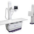
While the new generation of wireless DR detectors offers more flexibility than older systems designed to be installed in a room, these units are not yet fully portable, requiring separate devices, power cables, and data communication to function, the authors noted. At the same time, advances in radiography technology have resulted in smaller x-ray generators and other components that enable the development of mini-mobile DR systems that could use wireless "smart" devices such as phones and tablet computers.
The authors decided to explore the potential of mini-mobile DR by developing a prototype system designed for high portability, weighing less than 25 kg (55 lb) and measuring 66 x 45 x 33 cm (26 x 18 x 13 inches). The system employs a 120 x 150-mm flat-panel digital detector based on complementary metal-oxide-semiconductor (CMOS) technology, and an x-ray generator with a tube current ranging from 40 kV to 80 kV. The integrated x-ray generator and detector differentiate the unit from other small portable DR units, according to the authors.
For additional flexibility, the unit lacks a built-in display for viewing images. Instead, the system is designed to transmit images wirelessly from its embedded PC to external devices such as smartphones, tablet PCs, and iPads. Medical staff can view images with an Android-based application whenever they have network access, and the app has functions such as pan/zoom and measurement that are available when the user is offline. The app also enables users to employ their smart device as a remote control to operate the system.
For more rapid image review, raw image data are converted to JPEG files; images can be converted to DICOM format if they need to be transferred to a PACS, the researchers noted. Jeong and colleagues decided to test their unit by comparing its speed with that of their hospital's PACS when transferring images to mobile devices.
The mini-mobile DR system was able to transfer a variety of images in file sizes ranging from 46 KB to 281 KB at speeds ranging from 100 msec to 200 msec. Meanwhile, the PACS network took from 500 msec to nearly 800 msec to transfer images of the same size, making the mini-mobile DR a minimum of 400 msec faster.
"Smaller image sizes and faster response times mean less processing and transmission time required on the smart device, which leads to lower power consumption and a faster imaging service," the authors wrote. "This satisfies the physical constraints of smart devices."
The authors concluded by positing that the mini-mobile DR unit is a good solution for many clinical applications, such as for fracture patients, with greater flexibility and lower overhead than existing DR systems.
In future work, they plan to perform clinical evaluations and diagnostic tests of the system. They also plan to research the minimization of x-ray dose using the technology.


