Tomosynthesis with digital radiography (DR) could be a useful tool for following patients with neutropenia for signs of fungal pneumonia. Researchers from the University of Washington said the technique could offer better performance than conventional x-ray without the radiation dose of CT.
Neutropenic patients are frequently at risk for fungal pneumonia, which has a 10% mortality rate. Therefore, it is important to follow them with some kind of imaging modality, said Dr. Rachael Edwards, a radiology resident at the University of Washington Medical Center, in a talk at ECR 2014 in March.
Early detection of neutropenic pneumonia has been shown to reduce mortality, but the most commonly used screening modality -- chest x-ray -- has poor sensitivity, Edwards told attendees. CT has higher sensitivity but much higher radiation dose as well, which can be an issue when neutropenic patients are given weekly imaging studies to detect pneumonia.
Edwards and colleagues believe tomosynthesis could be a happy medium, with a lower dose than chest CT and sensitivity between radiography and CT.
Simulated tomosynthesis
One novel aspect of the study is its use of a simulated DR tomosynthesis protocol, in which tomosynthesis images were reconstructed from CT scans rather than from an actual DR system, according to Dr. Gregory Kicska, PhD, senior author on the study.
The software analyzes CT data to simulate the projections created from an x-ray source and an image detector, Kicska told AuntMinnie.com in an email, and it models mathematically what would be produced by an actual DR tomo unit. While the technique might not be optimal for clinical use, it can be a superior research tool as it enables patient databases to be mined for rare diseases, with CT data reconstructed to determine how tomosynthesis might affect care, he said.
For the retrospective study presented at ECR 2014, the researchers tested their hypothesis that tomosynthesis would be a better tool for surveillance using a group of 65 individuals seen at their institution. The study group consisted of 52 patients with early pneumonia and 13 who were normal.
Chest radiographs were performed within seven days of CT acquisition, and the CT scans were used to digitally reconstruct the simulated digital tomosynthesis images. Images were then rated on a 10-point scale by four cardiothoracic radiologists with minimal tomosynthesis experience. They graded images based on the size of fungal pneumonia nodules (with smaller nodules considered to be more difficult cases) and the likelihood of infection. The median nodule size was 18 mm.
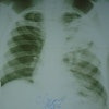
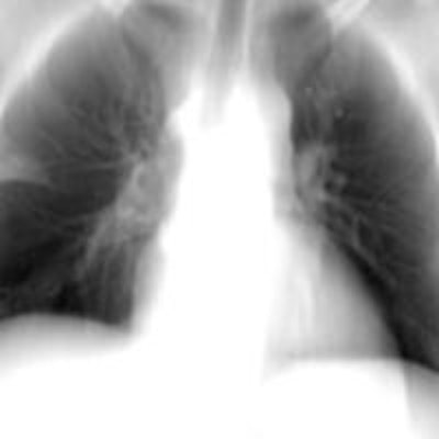
 Conventional chest radiograph (left) and simulated tomosynthesis image (right) for the same patient. Middle image shows a coronal CT slice that demonstrates early pneumonia. Note how this abnormality is clearly demonstrated in the tomosynthesis image but is more subtle in the radiograph. Image courtesy of Dr. Gregory Kicska, PhD.
The area under the curve (AUC) for the observers grading the nodules was 0.85 for tomosynthesis and 0.80 for conventional radiography, the researchers found. While the difference was not statistically significant, with a p-value of 0.28, the finding hints at a performance advantage for tomo that could be borne out in a real-world environment with actual DR tomo systems, according to Kicska.
"The fact that the AUC was better for tomosynthesis compared to radiography is very promising," Kicska said. "In the real world, tomosynthesis images are always accompanied by a traditional radiograph. Therefore, tomosynthesis in the real world would likely perform even better than our study suggests."
In fact, just a small increase in AUC would have a large clinical effect on the neutropenic population, he believes. In her ECR 2014 talk, Edwards presented several hypothetical scenarios that could demonstrate tomo's contribution to managing neutropenic patients at risk of pneumonia, based on the group's data.
For example, if both techniques performed at a desired specificity of 80%, tomosynthesis would have a sensitivity of 79%, compared with 70% for conventional radiography. In a patient population of 1,000 individuals screened for pneumonia, this means that tomosynthesis would diagnose 160 patients, compared with 140 using x-ray, avoiding 20 missed cases of pneumonia each year, Edwards said.
"With a mortality of 10%, this could be significant," she said.
Likewise, at a fixed sensitivity of 90%, tomosynthesis would have a specificity of 77%, compared with 58% for conventional radiography. In a hypothetical population of 1,000 individuals, the lower specificity means that 496 patients would proceed to CT for additional workup (160 positive cases and 336 false positives); meanwhile, with tomosynthesis, only 344 patients would need additional scanning (160 positive cases and 184 false positives) -- a difference of 152 individuals.
Kicska, Edwards, and colleagues plan to conduct additional research using an actual tomosynthesis system.
