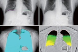
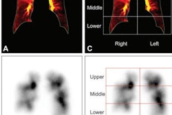
Konica Minolta Healthcare will showcase what it calls dynamic chest radiography as a work-in-progress at next week's annual World Congress of Thoracic Imaging (WCTI) in Boston.
Currently still in development, dynamic chest radiography uses Konica Minolta's flat-panel digital x-ray detector and proprietary data analysis software with the goal of enabling clinicians to simultaneously assess anatomical and physiological information, according to the vendor. As a result, a commonly performed chest x-ray could serve multiple functions and avoid subjecting the patient to multiple tests, Konica Minolta said.
The firm believes dynamic chest radiography could be used by clinicians to evaluate chest and lung function in patients with pulmonary diseases such as chronic obstructive pulmonary disease, asthma, and pulmonary embolism. Patient radiation exposure may also be reduced compared with the advanced imaging systems currently used for lung perfusion and ventilation studies, such as nuclear medicine and CT, Konica Minolta said.
A symposium on dynamic chest radiography will be held at WCTI and moderated by Dr. Heber MacMahon, section chief of thoracic imaging at the University of Chicago.


