Dear AuntMinnie Member,
A 3D software program developed at Stanford University in California is showing promise by creating fused PET/CT images that may improve clinicians' ability to find cancer.
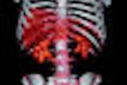
The Stanford researchers integrated PET's functional information with CT's anatomical information and presented it in a single 3D image with correct anatomic relationships, according to an article by staff writer Jonathan S. Batchelor that we're featuring in our Molecular Imaging Digital Community.
Using the 3D application they helped develop, the researchers conducted PET/CT scans on a variety of malignancies and analyzed the results. Waiting for the program to process the data took some patience, but the researchers found that the reconstructed images added valuable diagnostic information and changed patient management in several cases.
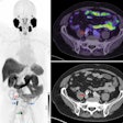
The article includes images as well as two short video clips that illustrate in stunning detail the potential of the technique in procedures such as virtual colonoscopy. To see them for yourself, just click here, or visit our Molecular Imaging Digital Community at molecular.auntminnie.com.


