Dear Molecular Imaging Insider,
Indium-111-labeled octreotide whole-body planar imaging has been a mainstay of nuclear medicine for detecting somatostatin receptor-positive neuroendocrine tumors. Most protocols call for imaging four to six hours and 24 hours after radiopharmaceutical injection. However, conventional modalities, such as CT or MR, are usually employed for more discrete anatomic localization in tracer uptake areas.
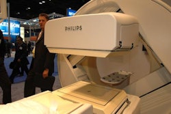
A research team from Ohio's Cleveland Clinic recently added SPECT/CT into the modality mix for octreotide imaging, and came up with some positive results that may encourage other clinicians to further investigate its possibilities.
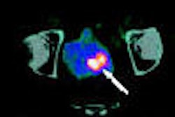
The scientists found that the utilization of SPECT/CT in octreotide imaging for neuroendocrine tumors provided important diagnostic information in nearly four out of five cases that was unavailable with planar whole-body scans. In addition, the use of SPECT/CT reduced false-positive results in almost 10% of the cases.
For more on how SPECT/CT is improving the diagnostic capabilities of octreotide imaging for neuroendocrine tumors, click here. As a Molecular Imaging Insider subscriber, you have access to this story before it's published for the rest of our AuntMinnie.com members.
Finally, if you haven't done so recently, be sure to take a look at our online reference book, Nuclear Medicine on the Internet. Dr. Scott Williams has updated chapters on myocardial perfusion exam findings, bone density measurement, I-131 therapy in thyroid cancer, various agents for tumor therapy, and a discussion on PET utilization for tumor imaging. Check out his most current postings by clicking here.


