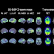
Researchers at the University of California, Davis recently tested the use of a total-body PET/CT scanner in COVID-19 patients, with results shedding light on the body's immunological response to the disease.
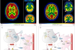
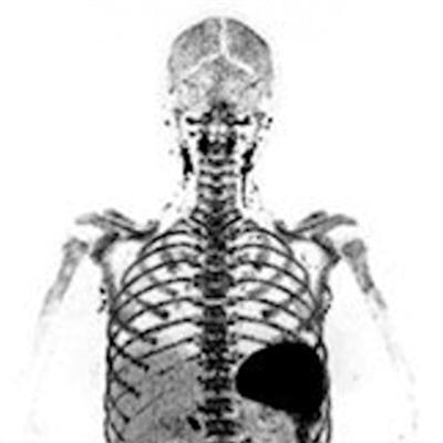
Biomedical engineering researcher Negar Omidvari, PhD, presented the images acquired with a total-body PET/CT scanner (uExplorer, United Imaging) at the Society of Nuclear Medicine and Molecular Imaging SNMMI 2022 annual meeting in June. The images show the distribution of CD8+ T-cells in recovered COVID-19 patients. Notably, the researchers used a new zirconium-89-based radiotracer designed to target T-cell activity.
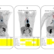
![The first reported image of CD8+ T-cell distribution in a recovered COVID-19 patient (a 29-year-old woman; body mass index [BMI] 29), imaged seven weeks post-symptom onset on a uExplorer total-body PET/CT scanner with a ~0.5 mCi injected dose of zirconium-89-Df-crefmirlimab-berdoxam radiotracer. Lymphoid organs containing CD8+ T cells (such as spleen, bone marrow, lymph nodes, and tonsils) showed the largest uptake, reflected by the darker black parts of the image. Image courtesy of Dr. Negar Omidvari at UC Davis.](https://img.auntminnie.com/files/base/smg/all/image/2022/08/am.2022_08_11_21_08_3322_2022_08_11_total_body_pet_resized.png?auto=format%2Ccompress&fit=max&q=70&w=400) The first reported image of CD8+ T-cell distribution in a recovered COVID-19 patient (a 29-year-old woman; body mass index [BMI] 29), imaged seven weeks post-symptom onset on a uExplorer total-body PET/CT scanner with a ~0.5 mCi injected dose of zirconium-89-Df-crefmirlimab-berdoxam radiotracer. Lymphoid organs containing CD8+ T cells (such as spleen, bone marrow, lymph nodes, and tonsils) showed the largest uptake, reflected by the darker black parts of the image. Image courtesy of Dr. Negar Omidvari at UC Davis.
The first reported image of CD8+ T-cell distribution in a recovered COVID-19 patient (a 29-year-old woman; body mass index [BMI] 29), imaged seven weeks post-symptom onset on a uExplorer total-body PET/CT scanner with a ~0.5 mCi injected dose of zirconium-89-Df-crefmirlimab-berdoxam radiotracer. Lymphoid organs containing CD8+ T cells (such as spleen, bone marrow, lymph nodes, and tonsils) showed the largest uptake, reflected by the darker black parts of the image. Image courtesy of Dr. Negar Omidvari at UC Davis.In the study, Omidvari and colleagues compared total-body PET/CT images between five recovering COVID-19 patients and three healthy volunteers, and they noted subtle differences in CD8+ T-cell deposition in the spleen, liver, bone marrow, lymph nodes, and tonsils of patients, as well as differences in the clearance rate of the radiotracer from blood.
"This research paves the way for further exploration of the assessment of immune response to infectious diseases and cancer, the assessment of response to vaccines in human subjects, and the fundamental mechanisms of the immune system in general," United Imaging said in a press release.


