Fibroblast activation protein inhibitor (FAPI) PET/CT appears superior to FDG-PET/CT for imaging gynecological cancers, according to a recent study.
The finding is from a head-to-head comparison between the two techniques for detecting primary tumors, lymph node metastases, and distant metastases, noted lead authors Tengfei Li, PhD, and Jintao Zhang, MD, of Southwest Medical University in Luzhou, China, and colleagues.
“[Gallium-68] FAPI-PET/CT demonstrated higher tracer uptake and was superior to F-18 FDG- PET/CT in detecting primary and metastatic lesions in patients with common gynecological malignancies,” the group wrote. The study was published February 28 in Cancer Imaging.
Studies indicate that breast cancer (11.6%), cervical cancer (3.3%), and ovarian cancer (1.6%) are the most commonly diagnosed gynecological malignancies and are leading causes of cancer deaths among women.
While F-18 FDG-PET/CT has proven valuable for the diagnosis, staging, and management of gynecological malignancies, it exhibits low sensitivity in detecting primary lesions and nodal metastases may not accurately differentiate between acute inflammatory infections and tumor growth, the authors explained. Conversely, experimental PET/CT studies with new FAPI radiotracers have shown significant tumor uptake and high sensitivity in head and neck, lung, gastric, colon, and esophageal cancers.
To further explore its potential, the researchers compared results among 35 patients with newly diagnosed or previously treated breast, ovarian, or cervical cancer who underwent paired F-18 FDG-PET/CT and Ga-68 FAPI-PET/CT scans to determine the most appropriate treatment strategy. Among the patients, 27 underwent imaging for the initial assessment of tumors, while eight were assessed for disease recurrence.
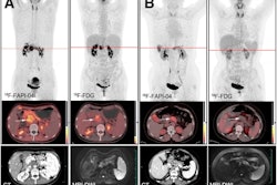
 A 47-year-old woman was admitted with a left breast mass. For staging, the patient underwent F-18 FDG-PET/CT. The maximal intensity projection image (A) and the axial views (B: PET image; C: CT scan; D: PET/CT fused image) revealed normal findings. Then she underwent Ga-68 FAPI-PET/CT. The left breast showed intense uptake on Ga-68 FAPI-PET/CT (E: PET image; F: CT scan; G: PET/CT fused image; H: MIP, solid arrow; SUVmax, 15.4). Subsequently, the patient underwent pathology confirmed invasive lobular carcinoma of the left breast. Image and caption available for republishing under Creative Commons license (CC BY 4.0 DEED, Attribution 4.0 International) and courtesy of Cancer Imaging.
According to the analysis, Ga-68 FAPI PET/CT was more sensitive than F-18 FDG PET/CT for identifying primary tumors in breast, ovarian, and cervical cancers (100% [27 of 27] vs. 85% [23 of 27], p < 0.001). Regarding the diagnostic performance for nodal metastasis, Ga-68 FAPI-PET/CT showed significant advantages in specificity (100% [35 of 35] vs. 66% [23 of 35], p < 0.001) and accuracy (94% [67 of 71] vs. 80% [54 of 71], p < 0.001), the group reported.
However, no significant difference was found between Ga-68 FAPI-PET/CT and F-18 FDG- PET/CT in sensitivity for detecting lymph node metastases (97% [35 of 36] vs. 86% [31 of 36], p = 0.125), the investigators noted.
Finally, in terms of diagnostic performance for distant metastases, Ga-68 FAPI-PET/CT demonstrated a higher radiotracer uptake in metastatic bone disease (maximum standardized uptake value [SUVmax]: 12.9 vs. 4.9, p = 0.036).
“Ga-68 FAPI is a novel and highly promising radiotracer for PET/CT imaging,” they wrote.
Ultimately, precise staging and lymph node detection are essential for the treatment and prognosis of cancer patients, and this study suggests the technique could help assist clinicians in formulating effective treatment plans, according to the team.
“Larger prospective studies are needed to confirm this,” it concluded.
The full study is available here.