A PET/MRI study found that androgen deprivation therapy used to treat prostate cancer causes progressive arterial inflammation, particularly in the carotid arteries, abdominal aorta, and iliac arteries, suggesting patients need closer cardiovascular monitoring and preventive treatment strategies.
- Study Design: Researchers tracked 43 prostate cancer patients with two F-18 FDG-PET/MRI scans over a median of 184 days to measure arterial inflammation.
- Key Finding: Androgen deprivation therapy (ADT) was associated with significant increases in arterial inflammation across multiple vascular territories, with the largest changes in iliac and carotid arteries.
- Progressive Risk: Arterial inflammation increased monthly by 0.0205 in iliac arteries and 0.0123 in carotid arteries, corresponding to cumulative increases of 0.15 to 0.25 over 12 months.
- Clinical Implication: The findings suggest cardiovascular risk assessment should be systematically integrated into care for all patients receiving prolonged ADT therapy.
- Future Direction: Researchers propose that statins and antiplatelet therapy may help mitigate arterial inflammation, though larger studies are needed to confirm preventive benefits.
F-18 FDG-PET/MRI shows that androgen deprivation therapy is associated with progressive arterial inflammation in men with prostate cancer, according to a recent study.
A group from Shanghai Jiao Tong University in Shanghai, China, quantified arterial inflammation across ADT exposure in patients using paired F-18 FDG-PET/MRI scans, with findings suggesting arterial inflammation may represent an imaging phenotype of ADT-associated cardiovascular risk, noted lead author Song Xue, PhD, and colleagues.
“These results underscore the need for systematic cardiovascular risk assessment and management in all patients receiving prolonged [androgen deprivation therapy],” the researchers noted. The study was published July 28 in Circulation: Cardiovascular Imaging.
Androgen deprivation therapy (ADT) is widely used in advanced prostate cancer and improves outcomes in patients, yet it is associated with increased cardiovascular events such as stroke and heart attacks, the authors explained. The underlying vascular biology driving the connection remains incompletely defined, they noted.
To address the gap, the researchers conducted a longitudinal study in 43 men with histologically confirmed prostate cancer. Participants underwent two whole-body F-18 FDG-PET/MRI scans (uPMR 790, United Imaging) at Renji Hospital across the course of ADT exposure. The enrollment period ran from May 2019 to December 2024, with a median interval of 184 days between scans.
Arterial inflammation was quantified by measuring F-18 FDG radiotracer uptake in the arterial wall relative to background activity in the blood pool (the maximum target-to-background ratio [TBRmax]) across the common carotid arteries, aortic arch, ascending and descending thoracic aorta, abdominal aorta, and common iliac arteries.
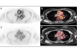
 Representative axial fused F-18 FDG-PET/MRI images and corresponding anatomic MRI images from a patient imaged at baseline and after initiation of androgen deprivation therapy are shown. Each row displays a distinct vascular territory. White arrows indicate focal areas of increased tracer uptake within the arterial wall, where spherical volumes of interest (VOIs) were placed for maximum standardized uptake value (SUVmax) measurement. Additional VOIs were placed in the superior vena cava and inferior vena cava for blood-pool activity sampling. SUVmax values from the arterial VOIs were extracted on the corresponding PET images and normalized to the blood-pool mean standardized uptake value to derive the maximum target-to-background ratio.Circulation: Cardiovascular Imaging
In secondary results, linear mixed-effects models showed progressive, time-dependent TBRmax rises, with the largest monthly increments in the iliac arteries (0.0205/month; p < 0.001) and carotid arteries (0.0123/month; p < 0.001), corresponding to cumulative increases of approximately 0.15 to 0.25 over 12 months. The abdominal aorta also showed a significant monthly rise (0.0098/month; p = 0.038), the researchers reported.
“In patients with prostate cancer, ADT is associated with consistent and progressive arterial inflammation, most prominently in carotid, iliac, and abdominal aortic regions,” the group wrote.
Ultimately, the longitudinal findings support a mechanistic link between androgen suppression and accelerated arterial inflammatory activity and raise the possibility that statin and antiplatelet therapy may mitigate the effect, the researchers suggested. Cardiovascular risk assessment may deserve closer integration into the care of patients receiving ADT, they wrote.
“Larger prospective studies are required to determine whether early preventive strategies aimed at reducing arterial inflammation can translate into lower cardiovascular event rates,” the authors concluded.
Read the full study here.