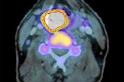
Researchers in Germany and the U.S. hoped that switching radiopharmaceuticals from FDG might improve PET's performance for imaging leukemia. While they found some benefits to using the new tracer, they also discovered some shortcomings.
The pilot study at the University of Ulm in Germany and the University of Iowa in Iowa City used the radiotracer 3'-deoxy-3'-18F-fluorothymidine (F-18 FLT) to image 10 patients with acute myeloid leukemia. The study was published by lead author Dr. Andreas Buck and colleagues in the November issue of the Journal of Nuclear Medicine (November 2008, Vol. 49:11, pp. 1756-1762).
The researchers were hoping that F-18 FLT might improve upon FDG, PET's workhorse tracer for oncology applications. FDG has been used for detecting hematologic neoplasms such as extramedullary leukemia based on their altered glucose consumption, but its utility may be reduced in areas of high natural glucose metabolism, such as the meninges or the pericardium. PET with FLT has the potential for use in these areas, Buck and colleagues wrote.
Patient population
The prospective study evaluated four men and six women with a mean age of 47 years, ranging from 23 to 70 years of age. Patients with high-risk acute myeloid leukemia (AML) scheduled for subsequent myeloablative treatment and bone marrow transplantation were eligible to participate in the study. Patients who had radiation or chemotherapy within four weeks were excluded from the study.
The researchers included 10 healthy control patients who were examined previously for further workup of indeterminate pulmonary nodules for reference. Standardized uptake values (SUVs) were calculated for reference segments of bone marrow, spleen, and normal organs.
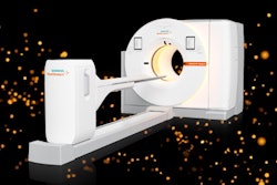
PET (ECAT HR1, Siemens Healthcare, Erlangen, Germany) was performed in five patients, producing 63 contiguous slices per bed position. The researchers noted that the axial field-of-view was 15.5 cm per bed position, with five bed positions for each patient, resulting in a total field-of-view of 77.5 cm.
The emission scan began 60 minutes after intravenous injection of approximately 370 MBq of F-18 FLT, which included the base of the skull, neck, thorax, abdomen, pelvis, and proximal femora in all 10 patients.
In the other five patients, images were acquired on a PET/CT system (Discovery LS, GE Healthcare, Chalfont St. Giles, U.K.). The CT acquisition protocol included a low-dose CT scan with a 0.5-sec rotation and 5-mm slice thickness. The scans were performed from the top of the skull to the midthigh for attenuation correction, followed by the PET scan acquired in 2D mode at three minutes and 40 contiguous slices per bed position.
The researchers found active AML disease in nine of 10 patients. Study results included one patient with a history of acute myelomonocytic leukemia because of clinical suspicion of relapse. Further analysis by bone marrow biopsy revealed a second complete remission. Two patients had refractory disease with leukemic blast infiltration of 80% and 100%, respectively.
Bone marrow SUV
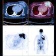
The analysis revealed that bone marrow was the organ with the highest uptake of F-18 FLT in all 10 patients with AML. Four patients presented with F-18 FLT uptake in the proximal femora and humeri not visible in the control group, which, the study stated, indicated bone marrow expansion.
Most notably, retention of F-18 FLT was observed predominantly in the bone marrow and spleen and was significantly higher in AML patients than in the control group. The mean F-18 FLT SUV in bone marrow was 11.5 for the AML patients and 6.6 in the control group, and the mean F-18 FLT SUV in the spleen was 6.1 in the AML patients and 1.8 in the control group.
Outside bone marrow, focal F-18 FLT uptake showed extramedullary manifestation sites of leukemia in four patients (meningeal disease, pericardial, abdominal, testicular, and lymph node), proven by other diagnostic procedures.
However, the researchers also found that there was no statistical significance between F-18 FLT uptake in normal bone marrow and the proliferative activity of leukemic blasts. This in particular can occur following treatment, which was the case in nine of 10 patients.
In conclusion, the researchers determined that the "preferential uptake of F-18 FLT in leukemia manifestation sites indicates that molecular imaging of leukemia based on deregulated cell cycle progression is feasible. Implications for clinical management and assessment of individual prognosis need to be addressed in a larger series."
By Wayne Forrest
AuntMinnie.com staff writer
January 7, 2009
Related Reading
PET dose-painting methods produce discordant results, August 27, 2008
Early PET scans may predict response to chemo in leukemia, July 29, 2008
Siemens makes FLT available to NCI research sites, November 21, 2007
Treatment-related AML and MDS rates 'acceptably low' after radiotherapy for NHL, October 5, 2007
Leukemia risk increases after breast cancer therapy in older women, September 25, 2007
Copyright © 2009 AuntMinnie.com


