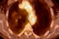
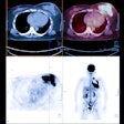
PET imaging of non-small cell lung cancer (NSCLC) prior to receiving radiation therapy should not be the basis for determining areas that may benefit from higher doses of radiation, according to research presented today at this week's American Society for Radiation Oncology (ASTRO) conference in Chicago.
The study from Thomas Jefferson University Hospital in Philadelphia was presented by Dr. Nitin Ohri, radiation oncology resident, who analyzed the PET scans of 43 patients. Fifteen of the 43 patients had significant activity on the scans both before and after treatment.
Ohri created a coordinate system that divided tumors into nine regions, or 17 regions for larger tumors. He then correlated the activity in the regions both before and after treatment.
Ohri found that in some patients, the activity pattern was in similar regions before and after treatment. However, there were some patients who showed activity in completely different areas after treatment compared to before treatment.
He said that it is not sufficient to increase the dose to areas that are especially active on PET imaging before treatment and expect it to improve the control rate, adding that it may be more appropriate to do a scan halfway through treatment and plan additional radiation dose around that scan.
Related Reading
PET shows promise for migraines, schizophrenia, October 30, 2009
UCLA study finds PET with FDDNP radiotracer can predict Alzheimer's, January 13, 2009
PET may help differentiate dementia syndromes, uncover ALS, December 2, 2008
PET, PiB support cognitive reserve hypothesis in Alzheimer's, November 14, 2008
WSJ: FDA supports imaging agents for Alzheimer's, October 24, 2008
Copyright © 2009 AuntMinnie.com


