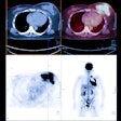
VIENNA - PET/MR is a feasible technique that yields diagnostic image quality in evaluating lung masses, according to a presentation at the European Congress of Radiology (ECR).
A team from Eberhard-Karls University Tübingen in Germany found that PET/MR produced similar lesion characterization and tumor staging to PET/CT in seven of 10 patients in their study.
"The results justify further investigation of MR/PET in the assessment of pulmonary lesions, especially with respect to the broad spectrum of function of MR techniques," said Dr. Christina Schraml. She presented the research during a Monday scientific session.
FDG-PET/CT is an established tool for routine staging and evaluation of therapy response in lung cancer. With the recent commercial availability of whole-body PET/MR systems, the researchers sought to compare the hybrid modalities in the assessment of pulmonary lesions regarding FDG uptake and tumor staging, Schraml said.
Ten patients (six males and four females) with highly suspected or known lung cancer were included in the study. Mean patient age was 61, with a range of 38 to 73.
All patients received a PET/MR scan directly after a PET/CT study. PET acquisition was performed after a contrast-enhanced whole-body CT scan, which was performed in the portal venous phase from the skull base to upper thigh.
In the PET/MR study, PET was acquired simultaneously to the MR sequences. The MR protocol on the 3-tesla scanner comprised an axial in-phase gradient echo (GE) sequence for attenuation correction and a coronal short tau inversion-recovery (STIR) sequence covering the lung and abdomen. In addition, a high-resolution axial T1 volume-interpolated breath-hold examination (VIBE) sequence of the lung was added, she said.
Patients received a mean dose of 347 MBq of FDG. The mean uptake time was 60 minutes for PET/CT and 126 minutes for PET/MR.
PET datasets from both modalities were co-registered using the TrueD software (Siemens Healthcare). Tumor to liver (T/L) ratios were computed by VOI analysis.
TNM classification staging was performed by two radiologists and one nuclear medicine physician in consensus. Of the 10 patients, eight had bronchial carcinoma and two had pneumonia.
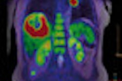
The researchers found that PET/CT and PET/MR exams were feasible in all patients. In addition, PET/MR showed diagnostic image quality without apparent image artifacts, she said. In nine of 10 lesions, PET/MR showed a pronounced FDG uptake.
At 8.0 ± 3.9, PET/MR had markedly higher T/L ratios than the 4.4 ± 2.0 produced by PET/CT.
"This was because the reference tissue, the liver, showed higher wash-out at the later time point," she said.
However, a strong Pearson's correlation (r = 0.90, p = 0.0008) was found between the T/L ratios on both scanners, Schraml said.
As for TNM staging, concordance between the two hybrid modalities was achieved in seven of 10 cases. Reasons for the discrepancies in the three cases included a mediastinal pleurae, a suprahilar lymph node, and one primary tumor size assessment, she said.


