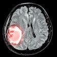
In the first study of its kind, researchers using 3-tesla MRI found a link between hippocampal volume, memory performance, and concussion severity among 28 former professional football players.
Texas researchers examined the hippocampus of retired NFL players, comparing them with a control group of 21 men of similar age and education. Primary investigator Dr. John Hart and colleagues collected high-resolution 3D images using a standard sensitivity encoding (SENSE) 8-channel head coil on a 3-tesla scanner (Philips Healthcare).
Imaging showed smaller hippocampal volumes among the retired players, who ranged in age from 36 to 79 years. Specifically, those with a history of concussion and loss of consciousness had smaller hippocampal volumes and lower memory test scores; however, the same relationship was not seen in those with a history of concussion who did not lose consciousness (JAMA Neurology, May 18, 2015).
Some of the players also met the criteria for mild cognitive impairment (MCI), which typically affects memory and may lead to dementia. The results were more pronounced among those who experienced more severe concussions, the researchers noted.
The results do not explain why the hippocampus was smaller in those who had more serious concussions, according to a press release from UT Southwestern Medical Center. A history of concussions and MCI appear to have a cumulative effect on hippocampal size and function, the group concluded.


.fFmgij6Hin.png?auto=compress%2Cformat&fit=crop&h=100&q=70&w=100)




.fFmgij6Hin.png?auto=compress%2Cformat&fit=crop&h=167&q=70&w=250)
