While painful and often debilitating for patients, migraines can also be difficult to diagnose and differentiate from other types of headaches. An MRI technique may help alleviate that diagnostic challenge, according to research presented on November 28 at the RSNA 2017 meeting in Chicago.
Dr. Melissa Meyer and colleagues from University Hospital Mannheim and Heidelberg University in Heidelberg, Germany, performed cerebral sodium MRI exams on healthy subjects and patients being clinically evaluated for migraine. They found that migraine patients had significantly higher sodium concentrations in the cerebrospinal fluid than those who did not have migraines.
"These findings might facilitate the challenging diagnosis of a migraine," Meyer said in a statement from the RSNA.
Affecting approximately 18% of women and 6% of men, migraines are one of the most common headache disorders. Migraines can cause severe head pain and sometimes nausea and vomiting; some may also cause vision changes or odd sensations in the body known as auras, according to the RSNA.
Unfortunately, the diagnosis is challenging because the characteristics of migraines and types of attacks vary widely among patients. As a result, many patients are undiagnosed and untreated, while other patients are given medications for migraines even though they have a different type of headache.
To help improve the diagnosis and understanding of migraines, the German researchers sought to evaluate the use of cerebral sodium MRI. Sodium has been shown in prior research to play an important role in brain chemistry, according to the society.
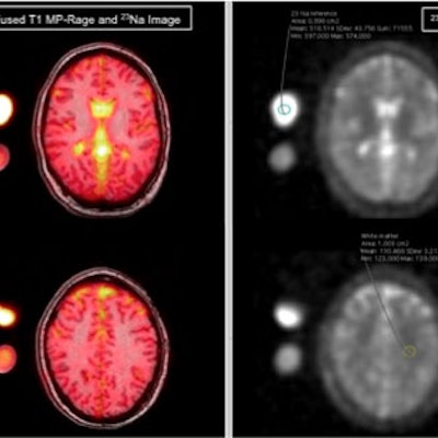
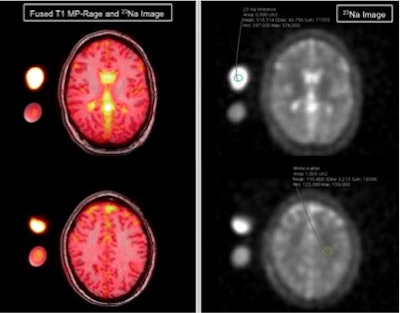 MRI examples of a migraine patient with exemplary region-of-interest placement in an external sodium reference phantom and in the white matter (right image). Left: fused T1-MP-RAGE and sodium image; right: sodium image. Images courtesy of RSNA.
The researchers recruited 12 women with a mean age of 34 who had been clinically evaluated for migraine. In addition, 12 healthy women of similar ages were added to the study to serve as a control group. The women who were being assessed for migraine filled out a questionnaire regarding the length, intensity, and frequency of their migraine attacks and accompanying auras. After both the migraine patients and healthy controls received cerebral sodium MRI exams, the researchers compared the sodium concentrations in both groups of women.
They found that migraine patients had significantly higher sodium concentrations in the cerebrospinal fluid, according to the RSNA. No statistically significant differences were found between the two groups in terms of sodium concentration in the gray and white matter, brain stem, and cerebellum.
The researchers said they hope to learn more about the connection between migraines and sodium in future studies.
"As this was an exploratory study, we plan to examine more patients, preferably during or shortly after a migraine attack, for further validation," Meyer said.


