WASHINGTON, DC - MRI scans with a diffusion-tensor imaging protocol (DTI-MRI) are helping to illustrate microstructural changes in the brain's white matter in people with cocaine use disorder, according to a study presented on Tuesday at the American Roentgen Ray Society (ARRS) meeting.
The findings from researchers at Florida State University (FSU) and Virginia Commonwealth University also provide new insights into potential peripheral markers of neuronal damage in certain brain regions, as well as the role oxidative stress plays in altering the structure of neurons and disrupting water flow and diffusivity.
"By using DTI and these peripheral markers of oxidative stress, we are able to detect damage far before there is ever a clinical presentation of psychiatric illness," said lead author Abbey Goodyear, a second-year medical student at FSU College of Medicine. "This could provide an opportunity for earlier diagnosis, as well as treatment."
Your brain on cocaine
Cocaine use disorder is associated with altered white-matter tracts and changes in microstructure in many regions of the brain. The brain region that's most consistently and notably mentioned is the corpus callosum, which is located beneath the cerebral cortex. The corpus callosum is associated with motor, sensory, and cognitive skills and integrates these functions on both sides of the brain, with the help of neurons that connect the two hemispheres.
 Abbey Goodyear from Florida State University.
Past studies have shown that changes in white-matter tracts due to cocaine use and other neurological disorders may be related to oxidative stress, which can be measured through DTI. The MRI technique, for example, has previously been used to evaluate oxidative stress in subjects with bipolar disorder.
DTI enables the visualization of changes in microstructure and white-matter tracts in the brain based on the diffusion of water molecules. With normal myelinated axons, the axial diffusivity is greater than the radial diffusivity because the flow of water moves forward from neuron to neuron. Axons are covered by a thick layer of myelin that prevents the diffusion of water in a peripheral direction.
"Healthy neurons have a nice layer of myelin, composed mostly of fat, surrounding their axons that provides insulation for the conduction of signals in the neuron," Goodyear explained. "Damage to these structures affects the way water diffuses through the brain, which can be analyzed through DTI."
When there is myelin damage, there is nothing to restrict the perpendicular flow of water, and the result is an increase in radial diffusivity. With acute axonal damage, axial diffusivity decreases due to fragmentation, which prevents the flow of water from an axon to the next neuron. With chronic damage, there is a general breakdown of the neuronal structure and an increase in both axial and radial diffusivity.
"In people with psychiatric illness or substance abuse disorder, there is the theory of oxidative stress," Goodyear told AuntMinnie.com. "This reaction compromises the structure of the fat in the brain, which surrounds the neurons and maintains their structural integrity. The oxidative stress alters the structure of the neurons, which disrupts the flow of water in the brain."
DTI analysis
To further investigate this connection, Goodyear and colleagues studied 28 subjects with cocaine use disorder (mean age, 35.4 ± 7.6 years) and 28 healthy, matched controls (mean age, 34.2 ± 8.4 years). Data on the cocaine users included alcohol use, current cocaine dependence, and whether their method of intake was nasal or through smoking.
Diffusion-tensor images were acquired on a 3-tesla MR scanner (Philips Healthcare). In all, 56 measures were obtained to test the relationship between oxidative stress and axial diffusivity, radial diffusivity, fractional anisotropy, and mean diffusivity. In addition, the International Consortium for Brain Mapping (ICBM) DTI-81 white-matter atlas was used to label brain regions that showed significant differences between the cocaine users and the healthy controls.
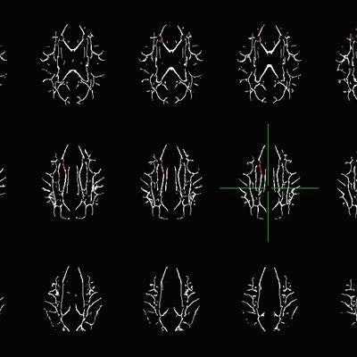
The most significant differences between the two groups were in axial diffusivity measurements (p < 0.05) in the anterior (or knee) of the corpus callosum, the body of the corpus callosum, the left superior corona radiata, and the left anterior corona radiata. The axial diffusivity values within these white-matter regions also correlated significantly (p < 0.05) with several oxidative stress measures. The researchers found no significant differences between the groups for fractional anisotropy, mean diffusivity, and radial diffusivity.
 Axial view of white-matter tracts in the brain. Computer analysis was used to determine statistically significant differences between those with cocaine use disorder and healthy subjects. Image courtesy of Abbey Goodyear.
"These findings strengthen the use of axial diffusivity as a measure of white-matter change proposed as axonal fragmentation and decreased connectivity, provide new insight into peripheral markers of neuronal damage in specific brain regions associated with cocaine use disorder, and provide further evidence for the oxidative stress theory of cocaine use disorder associated microstructural change," Goodyear said.
While the results of the study are promising, it is difficult to know how the findings might influence the treatment of cocaine use disorder. In general, the field of psychoradiology is still emerging, Goodyear said, but it does offer great potential for the early detection of psychiatric illness.
"Peripheral biomarkers are also being explored as inexpensive ways of detecting disease," she added. "Additionally, the pathophysiology of addiction continues to be studied and results seem to indicate targets for therapeutic intervention. It is impossible to say if our particular findings will one day be used clinically; however, I do believe our research may help move discovery in that direction."


.fFmgij6Hin.png?auto=compress%2Cformat&fit=crop&h=100&q=70&w=100)


.fFmgij6Hin.png?auto=compress%2Cformat&fit=crop&h=167&q=70&w=250)


