Researchers from South Korea have shown in a study published May 17 in Radiology that higher MRI volumes of a network of blood vessels in the brain called the choroid plexus are linked to more advanced Alzheimer's disease.
The findings suggest that choroid plexus volume and permeability may be potential imaging markers for Alzheimer's disease cognitive impairment, wrote a team led by Dr. Won-Jin Moon, PhD, of Konkuk University School of Medicine in Seoul.
"I think our findings on the choroid plexus can suggest it as a new potential MR imaging surrogate for an impaired clearance system and neuroinflammation," Moon said in a news release from the RSNA.
The choroid plexus is the primary site for the production of cerebrospinal fluid, which is crucial to clearing waste products and toxic proteins from the brain. There is mounting evidence that Alzheimer's disease is related to a failure in the brain to clear toxic proteins (namely, beta amyloid and tau), according to the researchers.
"We assume that the abnormal status of choroid plexus is linked to the failure of clearance, leading to waste and toxic protein accumulation in the brain and failure of immune surveillance leading to neuroinflammation," Moon explained.
To elucidate this issue, the group analyzed imaging from a group of 532 patients with various stages of cognitive impairment due to Alzheimer's disease. Seventy-eight patients had subjective cognitive impairment (SCI), 158 had early mild cognitive impairment (MCI), 149 had late MCI, and 147 had Alzheimer's disease.
Of these patients, they focused on 132 who underwent dynamic contrast-enhanced (DCE) imaging and quantitative susceptibility mapping (QSM) between January 2013 and May 2020 at Konkuk University Medical Center. Choroid plexus volume was automatically segmented using 3D T1-weighted sequences.
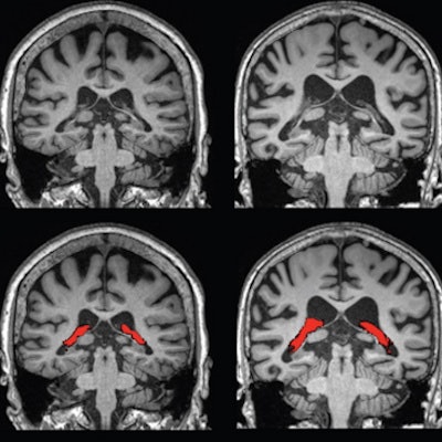
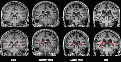
 Comparisons of four representative 3-tesla brain MRI scans of choroid plexus (CP) volume (red) according to disease stage over the cognitive impairment spectrum. CP volume is greater in the patient with Alzheimer disease (AD) than in those with subjective cognitive impairment (SCI) or mild cognitive impairment (MCI). All patients were 75-year-old women. Image courtesy of Radiology.
Results showed a stepwise increase in choroid plexus volume with severity of disease stage, with the SCI group having the smallest volumes and the Alzheimer's disease group having the largest. Moreover, the increase in choroid plexus volume was significantly associated with decreases in cortical and hippocampal volumes, which are more traditional markers of atrophy in Alzheimer's disease, according to the findings.
Importantly, the researchers found that larger choroid volumes were also associated with decreased memory, frontal executive function, and visuospatial function.
"Our study found that the enlarged choroid plexus volume is independently associated with increased cognitive impairment," Moon said. "We found no relationship between choroid plexus volume and amyloid pathology, but a clear relationship between the choroid plexus volume and cognitive impairment severity," he added.
The researchers concluded that further validation and longitudinal studies of choroid plexus volumes could lead to clinical methods for discriminating between vulnerable and less vulnerable patients.
In an accompanying editorial, neuroradiologist Dr. Gloria Chiang, an associate professor of radiology at Weill Cornell Medical College in New York City, suggested the study should be confirmed specifically in a prospective cohort with deep phenotyping for the pathologic features of Alzheimer's disease.
However, she echoed the potential clinical impact of the study.
"In the future, one can envision therapies that potentiate [choroid plexus] function in older age, leading to preserved [cerebrospinal fluid] secretion and potentially increased glymphatic flow to clear out toxic proteins," she said.
To get there, multimodal targeted imaging approaches to monitor choroid plexus function will be crucial, Chiang concluded.


