Diffusion-tensor imaging (DTI-MRI) has revealed new information about how the brain clears waste in patients with Alzheimer’s disease, according to a study published September 16 in Radiology.
The finding is from an analysis of postmortem human brains with and without Alzheimer’s disease and further supports the use of an emerging technique called DTI analysis along the perivascular space (DTI-ALPS), noted lead author Sihui Li, a doctoral candidate at Zhejiang University in Hangzhou, China, and colleagues.
“Diffusion tensor imaging (DTI) analysis along the perivascular space (ALPS) has been proposed to assess global glymphatic activity in several neurologic diseases. However, it is unclear whether this measurement is biased by fiber microstructure,” the group wrote.
The brain’s glymphatic system -- perivascular spaces along arteries and veins that serve as waste-clearing pathways -- has only recently been revealed on MRI but is an intense new area of research. DTI-ALPS provides a measurement (ALPS index) of fluid dynamics in the medullary perivascular spaces, with multiple studies reporting that the ALPS index is reduced in Alzheimer's disease, likely because of the buildup of beta-amyloid deposits, the authors explained.
However, it remains controversial whether the lower ALPS index in Alzheimer’s disease reflects glymphatic dysfunction or microstructural alterations, the group noted. To bridge the knowledge gap, the researchers conducted a DTI-ALPS analysis in a large cohort of older adults (18 donors with or without AD) and 110 participants from the Alzheimer's Disease Neuroimaging Initiative (ADNI).
The group first investigated the effect of microstructure on the ALPS index using DTI on the postmortem brains. The researchers demonstrated that microstructural asymmetry of white-matter fibers was not only evident in postmortem brain but was also age-dependent (r = −0.48; = p 0.044) and beta amyloid-status dependent.
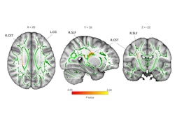
 A graphical abstract of the study. Aβ = beta-amyloid.RSNA
Next, to minimize microstructural effects, the researchers proposed a “corrected” ALPS index and further evaluated it in 110 participants from the ADNI. In contrast to the original ALPS index, the corrected ALPS index revealed no evidence of a difference between normal and clinical or pathologic Alzheimer’s disease groups and showed weaker correlations with clinical assessments in the ADNI dataset.
“The findings demonstrated that the ALPS index was partially driven by the microstructural asymmetry of the fibers,” the group wrote.
Altogether, the study indicates that microstructural asymmetry could be a useful biomarker of Alzheimer’s disease that is not at risk of being misinterpreted as glymphatic function, the researchers concluded.
In an accompanying editorial, Alex Rovira, MD, and Deborah Pareto, PhD, of University Hospital Vall d'Hebron in Barcelona, Spain, noted that the study highlights a central limitation of the ALPS index -- that it is influenced not only by perivascular fluid flow but also by the integrity and orientation of surrounding white matter.
“These considerations underscore the need for caution when interpreting ALPS values, particularly when used as a standalone marker,” they wrote.
Ultimately, the study makes a valuable contribution to the evolving field of brain clearance imaging and affirms the feasibility and clinical relevance of DTI-ALPS, Rovira and Pareto added. As insights into the glymphatic system deepen, imaging approaches must evolve accordingly, they wrote.
“With continued methodologic refinement and careful contextual interpretation, [DTI-ALPS] holds promise for elucidating glymphatic dysfunction across a spectrum of neurologic disorders,” Rovira and Pareto concluded.
The full study is available here.