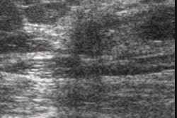
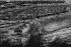
(Ultrasound Review) Microcalcifications demonstrated with ultrasound imaging could be successfully biopsied with sonographic guidance, according to researchers at Duke University Medical Center. Due to improvements in technology, now microcalcifications are more frequently visible with high-resolution ultrasound sonography.
"These microcalcifications are usually identified with hypoechoic masses that facilitate detection of the echogenic microcalfications," the authors reported.
When compared with stereotactic biopsy, ultrasound has certain advantages. There is no ionizing radiation involved; there is no compression of the breast; the patient positioning is more comfortable; and ultrasound guidance is faster and easier for the radiologist.
The study involved 23 patients that had microcalcifications detected on mammography. Initially 38 lesions were considered, but 40% showed no abnormality, uncertain findings or no microcalcifications on ultrasound imaging.
"Two groups of lesions were evaluated: microcalcifications within an associated mass or area of architectural distortion; and large clusters of microcalcifications with a diameter of 1 cm or greater containing numerous pleomorphic microcalcifications without an associated mammographic abnormality other than fibroglandular density," they wrote. The majority of lesions showed at least 15 pleomorphic microcalcifications at mammography, they said.
Patients either underwent percutaneous core biopsy with an automated gun and 14-gauge needle (69%) or 11-gauge vacuum-assisted technique (9%), or needle localization with surgical excision (22%). The authors reported "all 23 lesions were successfully biopsied under sonographic guidance with microcalcifications seen on specimen radiographs in each case." Further core biopsy and mammographic assessment of core was performed when no microcalcifications were shown on the first mammographic evaluation of the cores taken.
Pathological evaluation of malignant lesions showed ductal carcinoma in situ or invasive ductal carinoma, or both. They reported one benign lesion with microcalcifications that was a radial scar and ultrasound showed a dilated duct with echogenic foci. The authors suggested that further study is warranted to establish how sonographically guided evaluation of microcalcifications compares with stereotactic core biopsy.
"Sonographically guided biopsy of suspicious microcalcifications of the breast: a pilot study"M Soo et al
Department of radiology, breast imaging division, Duke University Medical Center, Durham, NC AJR 2002 April; 178:1007-1015
By Ultrasound Review
June 27, 2002
Copyright © 2002 AuntMinnie.com


