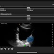
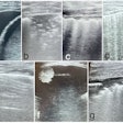
Ultrasound estimates of intramuscular fat can serve as a reliable indicator of physical health, according to research published online recently in Muscle & Nerve.
In a study of 42 participants with a wide range of body mass index (BMI) and physical fitness levels, a team from the University of Georgia found that an estimated percentage of intramuscular fat on ultrasound studies was inversely associated with physical activity and positively associated with body mass index.
"Ultrasound-estimated intramuscular fat was associated with other health measures and may provide physiological insight into the health consequences of obesity," wrote a team led by Dr. Hui-Ju Young.
After quantifying the estimated percentage of intramuscular fat estimates of four muscles (rectus femoris, biceps femoris, tibialis anterior, and medial gastrocnemius) using previously published equations, the group then compared the results with other health measures (Muscle & Nerve, March 11, 2016).
They found strong correlations in the percentage of intramuscular fat in the four muscles, with weak to moderate correlations between intramuscular fat and BMI, waist/hip ratio, muscle thickness, and muscle strength. They noted a significant relationship, however, between physical activity and intramuscular fat in the rectus femoris and medial gastrocnemius muscles (p < 0.05).
Ultrasound may be especially useful for providing muscle fat estimates in certain individuals who aren't well-suited for other imaging studies, according to Young. For example, those who have metal implants, muscle spasticity, morbid obesity, or pregnancy aren't a good fit with modalities such as dual-energy x-ray absorptiometry (DEXA), MRI, and CT.
"As research has supported the association between the amount of intramuscular fat and other health conditions, people with disabilities who have those conditions need a test that allows them to better understand their muscle composition," Young said in a statement.


