Digital mammograms may be best for informing radiomics models that assess breast density, according to findings published February 24 in Physica Medica.
A team led by Giacomo Feliciani from the Romagna Scientific Institute for the Study and Treatment of Tumors in Meldola, Italy reported that digital mammography outperforms synthetic mammography in depicting dichotomized breast density, yielding the highest informative content.
“Our results may be pertinent to the debate over screening mammography technique optimization using quantitative measures based on radiomics features,” the Feliciani team wrote.
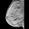
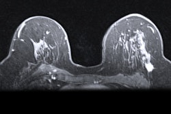
Breast density is a crucial aspect of risk assessment for the development of breast cancer in women, due to conventional screening mammograms struggling to detect cancers in dense breast tissue.
Researchers have pitted conventional mammography and digital breast tomosynthesis (DBT) against each other in studies to see which modality is best in finding cancers in women with dense breasts. Synthetic mammograms meanwhile are 2D reconstructions of DBT slices that some researchers say could eliminate the need for acquiring digital mammograms.
To better predict breast density, some researchers have turned to radiomics, where quantitative parameters are created from sets of images.
Feliciani and colleagues developed radiomics prediction models from both digital and synthetic mammograms. They investigated which breast images could best predict breast density in these models.
The team extracted a total of 123 imaging features from the 10 regions of interest of 96 women. After robustness analysis, it employed the most predictive features to build logistic regression-based models. The team developed radiomics models that used either digital mammograms, high-resolution synthetic mammograms, or standard synthetic mammograms.
The researchers compared the performance of each model with different bin sizes, bringing the total models developed to nine. These bins contain data points representing variables, used for image processing in imaging analysis. The bin sizes were 32, 64, and 128, respectively.
![Delineation workflow: The various steps of the contouring process: (a) the nipple is manually annotated (b) a reference point is placed 30 mm behind the nipple (c) 10 random theta/displacement values are selected to obtain the central point of each ROI (d) Regions of interest are placed on the image. (e) The resulting 10 ROIs obtained for a representative subject (viewing window [0 10000]). Images are published under a Creative Commons license (CC BY-NC-ND 4.0).](https://img.auntminnie.com/files/base/smg/all/image/2025/02/1_s2.0_S1120179725000523_gr1_lrg.67bcbbc3ac5a9.png?auto=format%2Ccompress&dpr=2&fit=max&q=70&w=700) Delineation workflow: The various steps of the contouring process: (a) the nipple is manually annotated (b) a reference point is placed 30 mm behind the nipple (c) 10 random theta/displacement values are selected to obtain the central point of each ROI (d) Regions of interest are placed on the image. (e) The resulting 10 ROIs obtained for a representative subject (viewing window [0 10000]). Images are published under a Creative Commons license (CC BY-NC-ND 4.0).
Delineation workflow: The various steps of the contouring process: (a) the nipple is manually annotated (b) a reference point is placed 30 mm behind the nipple (c) 10 random theta/displacement values are selected to obtain the central point of each ROI (d) Regions of interest are placed on the image. (e) The resulting 10 ROIs obtained for a representative subject (viewing window [0 10000]). Images are published under a Creative Commons license (CC BY-NC-ND 4.0).
The team found that the model using digital mammography data yielded the highest area under the curve (AUC) values in all three bin sizes.
| Comparison of digital, synthetic radiomics models AUCs for breast density prediction | |||
|---|---|---|---|
| Bin size | Standard synthetic mammography | High-resolution synthetic mammography | Digital mammography |
| 32 | 0.61 | 0.66 | 0.76 |
| 64 | 0.64 | 0.67 | 0.75 |
| 128 | 0.64 | 0.68 | 0.72 |
| *All data achieved statistical significance when compared to digital mammography. | |||
The study authors highlighted that their results shed light on how feasible it is to apply radiomics features to breast density prediction. They added that they could also identify the most appropriate imaging technique and quantization level for density prediction.
“As radiomics methodologies continue to improve, its integration into clinical practice holds the potential to enhance breast cancer risk prediction, ultimately benefiting patient outcomes,” the authors continued.
They also called for further research and validation in larger and diverse patient populations to confirm and extend the applicability of radiomics in breast imaging.
The full study can be found here.