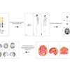
| FIGURE 1.1.3 Pedunculated advanced adenomas. 3D endoluminal CTC (A), coronal 2D CTC (B), and corresponding colonoscopy (C) images show a large pedunculated tubulovillous adenoma in the sigmoid colon. 3D endoluminal CTC image (D) from a second patie shows a 1.7-cm pedunculated polyp in the sigmoid colon, which is less conspicuous on the 2D CTC images (E, arrow) due to the similar appearance of hte sigmoid folds that are thickened by diverticular disease. The polyp was confirmed at same-day colonoscopy (F) and proved to be a tubulovillous adenoma. 3D endoluminal CTC (G) and double-contrast BE (H and I) images from three different patients show large pedunculated adenomas. |
Atlas of Gastrointestinal Imaging Figure 1.1.3 Pedunculated advanced adenomas
Latest in Home

Bone-RADS improves accuracy for junior, attending physicans
October 17, 2025


PET/CT reveals ‘chemo brain’ regions in leukemia patients
October 16, 2025


