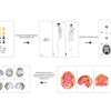
| FIGURE 1.1.5 Villous adenomas 3D endoluminal CTC (A) image shows a large, lobulated cecal mass. On prone 2D transverse CTC (B), the mass is submerged under the opacified fluid, which outlines its frond-like nature. The villous appearance is confirmed at colonoscopy (C). 3D endoluminal CTC image (D) from a second patient shows a relatively flat lobulated rectal mass (arrowheads), which carpets the mucosal surface. Note the rectal catheter (arrow. Images from subsequent colonoscopy (E) and EUS (F) show the same 5-cm mass. There is no evidence of submucosal extension at EUS, and this lesion proved to be a benign villous adenoma with high-grade dysplasia. 3D endoluminal CTC (G), 2D sagittal CTC (H), and colonoscopy (I) images from a third patient show another rectal villous adenoma manifesting as a flat carpet lesion, which involves a relatively large surface area and extends near the anorectal junction. |
Atlas of Gastrointestinal Imaging Figure 1.1.5 Villous adenomas
Latest in Home

Bone-RADS improves accuracy for junior, attending physicans
October 17, 2025


PET/CT reveals ‘chemo brain’ regions in leukemia patients
October 16, 2025


