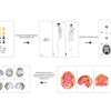
| FIGURE 1.1.6 Serrated adenoma. 3D endoluminal CTC (A), 2D transverse CTC (B), and colonoscopy (C) images show a large, drooping pedunculated mass near the hepatic flexure that proved to be a serrated adenoma at histologic examination. |
Atlas of Gastrointestinal Imaging Figure 1.1.6 Serrated adenoma
Latest in Home
Bone-RADS improves accuracy for junior, attending physicans
October 17, 2025


PET/CT reveals ‘chemo brain’ regions in leukemia patients
October 16, 2025


