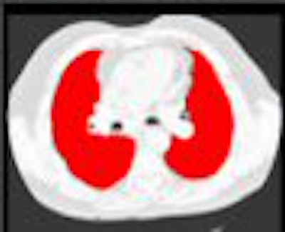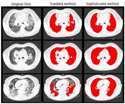
A lung segmentation algorithm that combines the advantages of standard and more sophisticated approaches yields fast and accurate results, according to researchers from the University Medical Center Utrecht in the Netherlands.
Automatic lung segmentation must be performed before computerized analysis of chest CT images, including computer-aided detection, can occur. However, standard lung segmentation algorithms may struggle when faced with large or subpleural abnormalities, according to Eva van Rikxoort, a doctoral candidate at the university's Image Sciences Institute.
"Important observations might be missed, or findings outside of the lung might be (taken into account)," she said.
More sophisticated algorithms come with their own problems, including computational complexity. To improve the situation, the Dutch researchers sought to develop a hybrid method that automatically detects a failure of the standard algorithm. When necessary, the hybrid approach can then resort to an advanced atlas-based method for proper segmentation, according to van Rikxoort.
"For clinical practice, there is always a compromise between speed and accuracy," she said. "This system solves that."
To test the method, the researchers employed 1,610 volumetric CT scans, including 1,360 scans taken from the NELSON Dutch lung cancer screening trial (30 mAs) and 250 scans from a database of patients with interstitial lung disease (50-120 mAs). Scans were performed using an Mx8000IDT or Brilliance 16 scanner (Philips Healthcare, Andover, MA) with 16 x 0.75-mm collimation, according to van Rikxoort. She presented the institution's findings at the 2007 RSNA meeting in Chicago.
The lungs in all 1,610 scans were automatically segmented with a fast algorithm, with errors automatically detected based on statistical deviation from a range of volume and shape measurements: left and right lung volume, ratio of volumes of both lungs, connected component analysis in 2D and 3D, and ratio between 2D lung volume in several slices, she said. Those scans with failures were then segmented using a multiatlas-based algorithm.
 |
| Image courtesy of Eva van Rikxoort. |
The segmentation algorithm automatically detected seven errors in the 1,360 screening scans, all of which were caused by pathologic abnormalities. All the errors were corrected by the atlas-based method, van Rikxoort said.
In 250 interstitial lung disease studies, 84 failures were automatically detected. Three scans were erroneously labeled as failures, and 81 were caused by pathologic changes. Of the 84 failures, the atlas-based method corrected 70 of 84 (83%).
The standard lung segmentation method took an average of 55 seconds, with error detection performed in an average of four seconds. The atlas-based method averaged 80 minutes.
For screening data, the average segmentation time was 79 seconds. Interstitial lung disease patients required an average segmentation time of 27 minutes. All results were handled on a single standard PC, van Rikxoort said.
"The new method combines the advantages of a fast and a more complex algorithm," she said.
By Erik L. Ridley
AuntMinnie.com staff writer
February 13, 2008
Related Reading
DR image processing produces mixed CAD results, February 12, 2008
Experience may make a difference with lung CAD, February 4, 2008
Radiologists more likely to reject certain true-positive CAD findings, January 22, 2008
CAD offers value in detecting lung nodules with CT, December 17, 2007
CAD may boost radiologists' ability to characterize lung nodules, November 25, 2007
Copyright © 2008 AuntMinnie.com


