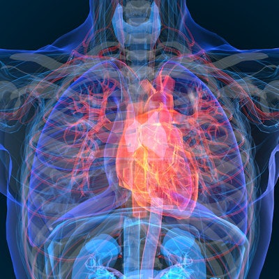
Radiomics analysis of low-dose CT lung screening exams can be used to stratify cardiovascular risk, creating a "one-stop" test for both lung cancer and heart disease, according to research published online June 30 in Academic Radiology.
Researchers from Rensselaer Polytechnic Institute (RPI) in Troy, NY, and Massachusetts General Hospital (MGH) found that prototype radiomics software could better differentiate patients with different Agatston and Multiethnic Scores for Atherosclerosis (MESA) risk scores than subjective assessment of coronary artery calcification and stenosis.
"With whole heart radiomics, LDCT can offer a 'one-stop' test for both lung cancer screening and cardiovascular risk stratification in at-risk subjects," wrote the researchers led by first author Dr. Fatemeh Homayounieh of MGH and senior author Dr. Mannudeep Kalra of RPI.
The researchers gathered data from 106 patients who had received LDCT for lung cancer screening at their institution between 2015 and 2019 and who had also undergone calcium scoring and coronary CT angiography (CCTA). All patients received their CT scans from one of three multidetector-row CT scanners from GE Healthcare, Philips Healthcare, and Siemens Healthineers.
Low-dose CT studies were performed at 120 kV, 30-50 mAs, 0.5-second rotation time, 0.9-1:1 beam pitch, 1-1.25 mm section thickness, and 0.8-1 mm section interval using vendor-specific iterative reconstruction, according to the researchers.
In addition to clinical variables, the authors recorded the subjective classification of coronary artery calcification, American College of Radiology Lung-RADS category, and presence of other pulmonary abnormalities for all LDCT exams. Agatston coronary calcium scores were recorded from the electrocardiogram (ECG)-gated calcium scoring CT, while the CAD-RADS classification for coronary artery stenosis was recorded from the ECG-gated coronary CT angiography study.
After the study subjects were risk-stratified based on their Agatston score and MESA 10-year coronary heart disease (CHD) risk score, prototype software from Siemens Healthineers was retrospectively applied to all 106 participants to automatically segment the heart and then perform radiomics analysis of the entire heart volume.
| Area under the curve (AUC) for radiomics analysis of LDCT exams | ||
| Clinical variables | Radiomics analysis | |
| Predicting extensive and nonextensive coronary calcifications | 0.69 | 0.92 |
| Predicting severe and nonsevere coronary artery stenosis | 0.69 | 0.86 |
| Predicting lower and higher MESA 10-year CHD risk groups | 0.79 | 0.87 |
The researchers also noted that whole-heart radiomics analysis better differentiated MESA 10-year CHD risks and the presence of significant coronary artery stenosis than the radiologists' visual assessment during LDCT interpretation. Also, combining LDCT-based subjective coronary calcification grading and whole-heart radiomics led to a slight improvement for prediction of high and low Agatston calcium scores from ECG-gated coronary calcium scoring CT, as well as for differentiating between lower and higher MESA 10-year CD risk groups.
The study reinforces the potential of cardiovascular risk assessment from LDCT exams performed for lung cancer screening, according to the researchers. With whole-heart radiomics, these exams can be used to screen for cardiovascular risks -- even without consideration of additional cardiovascular risk factors or subjective coronary calcification that's prone to interobserver variability and excludes extracoronary calcifications and epicardial fat, they said.
"Radiomics from LDCT can offer a 'one-stop' and cost-effective cardiovascular risk screening in at-risk subjects without the need for additional calcium scoring CT," the authors wrote.


