Convolutional neural network (CNN)-based AI image enhancement can render suboptimal chest CT images optimal, researchers have found.
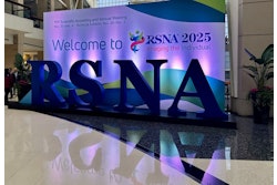
An added AI step could lead to salvaging unacceptable CT pulmonary angiography (CTPA) exams and performing fewer repeat imaging studies, according to Tejash Sikka, an Albany Medical College student who presented the project at RSNA 2025, on behalf of the Webster Center for Quality and Safety at Massachusetts General Hospital and Harvard Medical School.
During the COVID-19 pandemic, "a substantial number of CTPAs were rendered suboptimal due to inadequate contrast enhancement, often caused by large body habitus, cardiac output variability, and contrast timing issues," Sikka explained.
Aiming for improved contrast visibility and noise reduction, Sikka and colleagues applied AI-image enhancement to a total of 611 optimal and suboptimal CTPA and contrast-enhanced chest CT studies to determine the impact of the AI on image quality and diagnostic acceptability.
For 318 cases that thoracic radiologists identified as suboptimal due to insufficient vascular opacification or excessive image noise, Sikka noted that the AI improved streak artifacts and beam hardening artifacts.
Quantitative metrics Sikka noted included vascular attenuation values (HU) in the main pulmonary artery, ascending thoracic aorta, and segmental pulmonary arteries, as well as signal-to-noise ratios (SNR) and contrast-to-noise ratios (CNR).
HU values were measured from CT images with a standardized 1-mm thickness. Statistical analysis utilized paired t-tests comparing pre- and post-AI enhancement metrics.
After applying the AI, researchers observed a significant increase (p = 0.001) in attenuation pre-AI compared to post-AI: from 192 to 293 HU in the main pulmonary artery, from 235 to 362 HU in the ascending thoracic aorta, and from 187 to 282 HU in segmental pulmonary artery, according to the results.
Sikka also noted "massive" improvements in noise reduction: in the main pulmonary artery, from 17.6 HU to 11.2 HU; in the ascending aorta, from 17.2 HU to 10.8 HU; and in the segmental pulmonary arteries, from 19 HU to 14.7 HU.
Likewise, the group observed marked improvements in SNR and CNR, post enhancement, with all comparisons statistically significant (p = 0.001).
| SNR improvement | Pre-AI | Post-AI |
| Main pulmonary artery | 20.8 | 59.7 |
| Ascending aorta | 25.2 | 72.7 |
| Segmental pulmonary arteries | 20.1 | 56.9 |
| CNR improvement | Pre-AI | Post-AI |
| Main pulmonary artery | 188.8 | 288.8 |
| Ascending aorta | 232.1 | 357.3 |
| Segmental pulmonary arteries | 184.5 | 277.9 |
"This is the preliminary study where we are seeing if the AI works," Sikka noted. "We are working on other AIs that detect [pulmonary emboli] PEs in arteries automatically. If we have suboptimal images, using this AI we can enhance that image and then we can use that AI on top of the AI, a dual AI model, to make sure that PEs aren't missed."
Applying AI-based image enhancement in routine clinical practice can effectively salvage suboptimal chest CT studies, reducing repeat imaging, avoiding additional radiation and contrast exposure, and enhancing diagnostic confidence and accuracy in patient care, Sikka and colleagues concluded.
Next steps for the research will explore AI for distinguishing between artifacts and PEs, Sikka said.