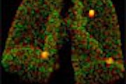
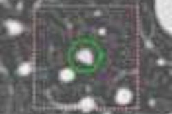
(Radiology Review) Computer-aided detection (CAD) enabled improved detection of lung nodules on chest radiographs, according to Japanese radiologists. Researchers at the University of Occupational and Environmental Health School of Medicine in Iseigaoka evaluated the use of a new commercially available CAD system (EpiSight/XR) that performs automated detection of lung cancer nodules.
Screening chest radiographs are extensively used for early detection of lung cancer, the number one cause of cancer-related deaths in the world. Improved diagnostic accuracy is essential according to Dr Shingo Kakeda and colleagues, writing in the American Journal of Roentgenology.
"The purpose of CAD is to direct the radiologist's attention by identifying and indicating suspected focal opacities that may represent cancer on a radiograph. Several previous studies have reported that CAD systems with automated detection methods for lung nodules could significantly improve diagnostic accuracy for identifying lung nodules on chest radiographs," they said.
Forty-five patients with a single lung nodule less than 25 mm diameter were compared with 45 normal subjects. Eight radiologists interpreted initial chest radiographs and CAD output images, and observer performance studies also were evaluated. "Individually, the use of CAD output images was more beneficial to radiology residents than to board-certified radiologists," they found.
False-positive CAD detections were prevalent with 315 false-positive detections on 100 normal radiographs. The majority (75%) of lesions was easily dismissed as normal anatomy, and 5% were outside the lung fields.
"Although the results of our observer study showed a significant improvement in the accuracy of lung nodule detection with CAD output images, our CAD performance level of 73% sensitivity is still not adequate to avoid overlooking lung nodules," they reported. Further improvement was necessary to improve the sensitivity, they asserted.
Temporal subtraction is another CAD technique that subtracts a previous chest radiograph from the current radiograph in order to enhance time-related changes. This method is useful if previous imaging with a similar projection has been performed.
Kakeda concluded, "this CAD system for digital chest radiographs can assist radiologists and has the potential to improve the detection of lung nodules due to lung cancer." By combining the automated lung nodule-detection method and temporal subtraction technique, the diagnostic accuracy of CAD might be further improved, they suggested.
Improved detection of lung nodules on chest radiographs using a commercial computer-aided diagnosis system
Kakeda, Shingo, et. al.
Department of radiology, University of Occupational and Environmental Health School of Medicine, Iseigaoka, Japan
AJR 2004 January; 182:505-510
By Radiology Review
March 18, 2004
Copyright © 2004 AuntMinnie.com

