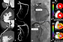
A machine learning (ML) model including both coronary CT angiography (CCTA) and stress cardiac MRI data can accurately predict major adverse cardiovascular events (MACE), suggest findings published January 14 in Radiology.
Researchers led by Théo Pezel, MD, from Université Paris Cité in France found that their model performed better than traditional methods and existing scores in patients with newly diagnosed coronary artery disease.
“This ML approach outperformed individual clinical, CCTA, and stress cardiac MRI metrics alone, as well as existing scores and traditional statistical methods, in two separate external validation datasets,” Pezel and co-authors wrote.
While current guidelines recommend CCTA as a noninvasive tool for diagnosing coronary artery disease, stress cardiac MRI has emerged as a more accurate risk stratification tool. The researchers highlighted that while multimodality imaging could be even more useful in this area, such an approach has been underused due to challenges in unifying data from different techniques at the same time.
The Pezel team developed and tested an ML model that uses data from both MRI and CCTA to predict MACE in patients with newly diagnosed coronary artery disease.
To develop the model, the team evaluated 18 clinical, two electrocardiogram, nine CCTA, and 12 cardiac MRI parameters as inputs. This involved automated feature selection with the least absolute shrinkage and selection operator (LASSO) algorithm and model building with an XGBoost algorithm.
The researchers also employed the model for training, testing, and validation of datasets from three institutions: The first was a training dataset of 1,427 patients and a testing set of 611 patients from Jacques Cartier Private Hospital; the second was an external validation dataset of 274 patients from Lariboisière Hospital; and the third was an external validation dataset of 269 patients from the American Hospital of Paris.
The retrospective study included data collected between 2008 and 2020 from 2,210 patients who completed cardiac MRI. Of these, 2,038 completed follow-up exams at a median duration of seven years. Of the total patients, 281 (13.8%) experienced MACE.
The ML model achieved a higher area under the curve (AUC) than traditional risk scoring and predictive methods.
| Performance of machine-learning model, traditional methods in predicting MACE in patients | |
|---|---|
| Method | AUC |
| Machine-learning model | 0.86 |
| European Society of Cardiology scoring | 0.55 |
| QRISK scoring | 0.6 |
| Framingham Risk Score | 0.5 |
| Segment involvement score | 0.71 |
| CCTA data alone | 0.76 |
| Stress cardiac MRI data alone | 0.83 |
| *p values ranged between < 0.001 and 0.004 for methods compared to the ML model’s performance. | |
The model also achieved superior performance in the two external validation datasets. These included AUCs of 0.84 and 0.92, respectively.
Finally, the team reported that the prevalence of inducible ischemia (15%), late gadolinium enhancement (11%), and annual rates of MACE (2%) were consistent with those of other stress cardiac MRI study populations.
The authors highlighted that ML approaches with multimodality imaging could be a precise method for predicting MACE with these results in mind. They called for prospective multi-center studies to be managed.
“ML methods are widely used and efficient for analyzing large amounts of data, whereas traditional algorithms falter because of inadequate model complexity,” the authors wrote. “For that reason, ML seems well suited for the comprehensive analysis of complex imaging datasets.”
However, they noted that the clinical application of such models may be limited due to the inherent challenges of performing both CCTA and cardiac MRI in the same patient.
The full study can be found here.


