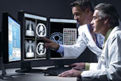
With more than 8.8 million exams stored in the PACS archive of Massachusetts General Hospital (MGH) in Boston, there's probably an image representing any medical condition and diagnostic interpretation imaginable. The challenge for radiologists or researchers is how to find it.
Render, a radiology report and image search application developed at MGH, makes it possible. This online searchable radiology study repository is described in the September issue of RadioGraphics (2009, Vol. 29:5, pp. 1233-1246).
What is it?
Render utilizes a database of all radiology reports contained in the hospital's information system since 1988. This database is continuously refreshed, with approximately 2,200 reports added each day. The content includes patient demographics, inpatient/outpatient status, unique exam number, and the text of the complete report.
Natural language processing software (Leximer, Nuance Communications, Burlington, MA) automatically analyzes the text content of both free text and structured reports. It identifies reports by positive and negative findings at a documented 97.5% accuracy level. Identifying whether a report contains positive (approximately 68%) or negative (approximately 32%) findings helps improve the relevancy of search results, according to lead author Dr. Pragya Dang.
All information contained in the reports is extrapolated and indexed by pertinent information categories, such as patient demographics (age and gender), exam information (date, type of procedure, modality, etc.), and positive or negative findings. The background indexing makes rapid, real-time searches possible.
Images that represent radiology exams of interest or that would be good teaching examples are selectively added to the 3-TB storage archive -- typically, 100 exams are added daily. While it is possible to access any image in the vast PACS archives, the subset of more than 1.6 million images contained in the Render archive meets most users' requirements.
Adding images
Images are acquired from PACS diagnostic workstations in one of three ways:
- Exams that have been viewed a total of three times by different radiologists or residents are added because they are likely to contain interesting findings or have the potential to be a good teaching exam.
- Radiologists may select individual images within an exam representing key findings -- or an entire exam -- for inclusion in the database.
- Images saved and exported in JPEG format for publication or teaching use by individual radiologists are automatically added.
DICOM images are automatically converted into an 8-bit JPEG format to reduce storage requirements and to make loading and viewing time faster for Render users. These are initially transferred from a PACS diagnostic workstation to a network-attached storage device that adds a unique examination number for each JPEG image, which also links it to the original exam.
Users may access the PACS archive to retrieve images not contained in the Render archive by launching the hospital's enterprise Web-based PACS. Selected images may be transferred into the Render network-attached storage, converted to JPEG format, and added to the Render archive.
Conducting searches
Two search modes are available, one for research applications and the other as a Web-search mode with six options.
The research mode displays results in a tabular format that includes categories for patient demographics (age, gender, and date of birth), exam information (date of exam, unique examination number, medical record number, and description of examination), imaging information (body region imaged and type of modality), radiology report contents, and images associated with the report. The information is provided in a format whereby it can be exported to a spreadsheet for analysis. If a user needs to view patient identifiers, institutional review board approval is required.
The Web-search mode offers a variety of search options to users. When a query term is used to conduct a search, Render's search engine retrieves all relevant cases with their reports and images for the query term, and for any synonym identified by a medical thesaurus file. Reports that are retrieved by the search display a portion of the radiology report and thumbnail images. When an image or report is selected, the entire file opens. The retrieval process for a basic query term search takes less than five seconds, according to the authors.
Selection of the full search option adds a significant amount of viewable detail to every entry. The additional detail includes a brief patient history or clinical indication and a complete list of all diagnostic imaging procedures that the patient has had.
An external search enables users to conduct Internet searches. A tagging feature lets users create a public or private bookmark, and tagged cases can be rapidly retrieved. Free-text searches may also be conducted.
Users who wish to conduct a very precise search can select the advanced search option. This option enables users to select specific fields in combination, such as type of modality, patient age and gender, exam date range, body regions, and single words or a combination of words contained in the clinical impressions or report text. Entries may also be selected in combination with other fields to identify reports associated with specific radiologists or that exclude negative findings in the reports.
By Cynthia E. Keen
AuntMinnie.com staff writer
November 23, 2009
Related Reading
New search tool finds the needle in PACS haystack, May 1, 2009
Language processing technology guides pathology, imaging studies, January 24, 2008
New tool automates unstructured report analysis, February 18, 2005
Copyright © 2009 AuntMinnie.com


