

 |
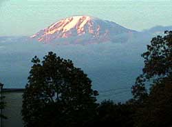
| The striking backdrop of Mt. Kilimanjaro as seen from the courtyard in front of the hospital. |
Thorough planning and plenty of descriptive information cannot begin to prepare a visiting radiologist for their first day at the Kilimanjaro Christian Medical Center in Moshi, Tanzania. The fatigue of the 17-hour flight contributes to the almost surreal experience of seeing this large African medical center, and case after case of incredible pathology, while trying your best to communicate with patients in a vestigial form of Swahili learned on the flight over.
During my visit, the first morning case conference consisted of a didactic review of approximately 15 cases (13 of them abnormal) obtained the night before, with such pathology as ankylosing spondylitis, osteomyelitis, abundant TB, and a right MCA stroke in a 37-year-old female who suffered this untoward event while climbing Mt. Kilimanjaro.
During the first pass through the department, two young children with Burkitt's lymphoma and large mandibular masses and one infant with clinically obvious hydrocephalus were waiting for ultrasounds while clinicians were busily attempting to obtain an echocardiogram on a young boy with severe post-infectious manifestations of rheumatic fever. The entire day's work consisted of 45 ultrasounds, 70 radiographs, and 5 CT examinations. Nearly all of the cases demonstrated some type of pathology, and nearly all of the patients, even those who were significantly debilitated, walked down to the department for their examinations. Although most of the patients had advanced disease they were unfailingly cooperative and very polite.
KCMC and its radiology department
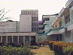 |
| The Kilimanjaro Christian Medical Center, a large open-air hospital with courtyards full of tropical foliage and wards replete with exotic disease. |
The Kilimanjaro Christian Medial Center (KCMC) is a 500-bed medical center located adjacent to Mt. Kilimanjaro and near the Serengeti Plain. It was originally constructed by missionaries in 1971 and is currently governed by a religious organization, the Good Samaritan Foundation.
The hospital is a referral center for East Africa, and patients come from hundreds of miles to receive medical care at the KCMC. The hospital itself is extremely busy and maintains an occupancy that constantly exceeds its bed capacity. Although the most common language spoken in the hospital is Swahili, all of the medical personnel speak English and all of the hospital records, including the radiology reports, are in English.
The operation of the radiology department is fueled by the endless energy of Dr. Helmut Diefenthal, a German-born U.S. citizen who has been involved with medical missionary work since 1956. Dr. Diefenthal first arrived in Tanzania (then Tanganyika) with his wife (Rotraut) in 1960 and practiced medicine until 1965, when he ventured to the U.S. and the University of Minnesota to pursue a residency in radiology.
After a two-year return to Tanzania to work at newly constructed Kilimanjaro Christian Medical Center, he went back to the U.S. to work at the University of Minnesota and the Veterans Administration Hospital. Following this 16-year hiatus, taken to put his four children through college, Dr. Diefenthal then returned to Tanzania and now functions as the driving force of the department of radiology. As such, he is intricately involved in many administrative components of the medical center.
The head of the department is Dr. Alpha P. S. Lyimo, a Tanzanian-born, Cuban-trained radiologist whose primary focus is plain-film radiography, GU, and GI work. In addition to Drs. Diefenthal and Lyimo, there are four radiology residents and seven assistant medical officers. The remainder of the staff consists of four radiologic technologists, one nurse midwife, one transcriptionist, and four office attendants.
The Kilimanjaro School of Radiology also teaches Tanzanian Assistant Medical Officers (AMOs) in radiology. Despite having 32 million people, Tanzania only has nine radiologists and about 1,300 physicians (about four physicians per 100,000 compared to 199 per 100,000 in the U.S.). The purpose of the AMO is, therefore, to provide some type of medical care for the country's significantly underserved population. Those accepted for the program already have three years of formal medical training, five years of practical work, and two additional years of training. The radiology program for AMOs involves two years of training in the basic sciences, radiation physics, film interpretation, and ultrasound.
The radiology residency at KCMC is a traditional four-year training program similar to training programs in the U.S. The curriculum is very similar to U.S. training programs, and excellent didactic conferences are given daily by Dr. Diefenthal, who does the vast majority of the resident teaching. Although there is extensive training in plain-film radiography, ultrasound, and computed tomography, they do not receive adequate experience in interventional radiology, nuclear medicine, and magnetic resonance imaging. This situation is a reflection of the state of radiology in Africa, where this training is not available because the appropriate equipment and the means to pay for it simply do not exist.
The department has three old but functioning radiographic units (a Philips Basic Diagnost, a Philips Compact Diagnost, and a GE Fluoricon) and a non-functioning fluoroscopy unit (a Philips Diagnost 5). The portable radiographic unit is a relatively modern Philips Practix 100, but the mammography unit is old (a CGR Senographe 500T) and is not in working order. The ultrasound machines vary, from the adequate Hewlett-Packard Sonos 100CF to a museum-candidate Medison scanner.
Surprisingly, the CT scanner is a new Philips Tomoscan SR 4000 with helical capability and many advanced features. This unit was purchased by the Tanzanian government and has proved to be not only a valuable diagnostic tool but, according to Dr. Diefenthal, "somewhat of a white elephant." By this he means that although it is valuable to have, the expense of maintaining it and the price that must be charged per scan is on the verge of being out of the range of affordability for even the most affluent Tanzanian patients.
Unlike in developed nations, two of the major barriers against the presence of advanced radiologic equipment are the ability to afford the technology and the lack of appropriate maintenance service. Both the fluoroscopy and mammography units could be promptly repaired if the appropriate parts and maintenance expertise were available. The charge for a CT scan of the head seems relatively low, at $70.51, but consider that this is approximately 70% of the monthly salary of an attending specialist physician working at the Medical Center. Additionally, the phenomenon of health insurance is all but unheard of, and nearly all medical bills are out-of-pocket expenses for the vast majority of Tanzanian citizens.
The daily work is also replete with numerous cost-saving measures that would be considered dramatic by Western standards. The latex gloves are all cleaned and reused. The sheets and pillowcases covering the ultrasound table are left in place for a week and are visibly very soiled by the end of this time period. The primary ultrasound scanning room is divided into two areas by wires and bed sheets hung from the walls, and Dr. Diefenthal's office is used as an additional ultrasound scanning room during the day.
There is a lack of type-print quality paper, and the official radiology reports consist of hand-written reports done in duplicate with the aid of carbon paper. Even pens and pencils are in very short supply, and radiographic film markers are nonexistent. Ultrasound images are printed on paper, and only images demonstrating some sort of pathology are captured. Only one film jacket per patient is used, with the numerous studies separated by a paper marker reused from other very worn film folders. Despite these substantial obstacles and incredible cost restraints, Dr. Diefenthal and his staff use a combination of resourcefulness and tenacity to consistently provide high-quality imaging services to the patients at KCMC.
The Diefenthals
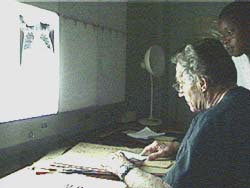 |
| Dr. Helmut Diefenthal in consultation with a pediatrician colleague discussing an infant suspected of having congenital heart disease. In addition to his expertise in general radiology, Dr. Diefenthal is well-known for his expertise in echocardiography. |
From a personal perspective, Dr. Diefenthal is one of those rare individuals who possess a strong combination of both dedication and altruism. He works very long days, teaches residents, performs examinations, and is constantly relied upon for his expertise and professional opinions. Despite his extremely demanding schedule, he works without salary and personally finances most of the required fees for the students through a six-week annual visit to the U.S., where he works as a radiologist at the University of Minnesota and at a private clinic.
He also heavily subsidizes the school fees and living expenses of one resident, providing financial support for his food, lodging, and textbooks. He and his wife Rotraut subsist on income from a retirement pension and live very modestly while providing invaluable assistance and incredible generosity to both visitors and permanent departmental staff.
The life of Helmut Diefenthal has been crammed full of enough interesting and incredible experiences for at least three people. Born and educated in Berlin, he was taken from medical school at the age of 18 and drafted into the German army as a medic. After an abrupt indoctrination into the military he was promptly sent to the Russian front for two years, where he was in charge of the Typhus control program. More dangerous than the front lines, this position had a mortality rate of 100% up to the time of Private Diefenthal's arrival, and all of his predecessors had died of the disease. Incredibly, while the vector for this disease is the louse, there were no delousing agents available and the lice ran rampant throughout the unit.
Amazingly, he survived this squalid existence and amidst the post-war rubble began medical school at the University of Berlin in 1945. Graduating in 1949, he began additional training in internal medicine and practiced at the university on staff until 1956, when he decided to become a medical missionary and was sent to Ipoh, Malaysia on a mission sponsored by the Lutheran Church in the U.S.
He spent four years in Malaysia as a general physician treating diseases typically seen in tropical climates. At that particular time the people in the country had great difficulty with Ancylostoma duodenale (hookworm) infections, and although there was treatment available, little to no information was available regarding the prevention of the disease (i.e. wearing shoes while walking outside). The policy of the Malaysian government was to deny the existence of the problem, and ideas to the contrary were not well accepted.
During Dr. Diefenthal's fourth year there, a news reporter contacted him about this endemic infectious problem and asked what could be done to ameliorate the situation. After his factually correct and straightforward answers were published, the Malaysian government expressed its outrage and the Diefenthals were subsequently forced from the country.
After a sabbatical in the U.S., Dr. Diefenthal was transferred to Tanzania, where he stayed in a remote hospital until 1965, when he returned to America and began radiology training at the University of Minnesota at the age of 41.
During this time, Ro Diefenthal gave up her Ph.D. studies in literature to become a radiologic technologist, studying at the University of Minnesota. This was not an easy decision, but she selflessly reasoned that this training would best prepare her for a vital role in the team effort of missionary radiology. Much like Helmut, Ro Diefenthal is extraordinarily bright, with a good sense of humor, and she possesses incredible altruism. Born in East Prussia, she epitomizes the hard work and dedication that characterized that region of German-speaking people. In addition to her native tongue, she speaks at least three other languages (that she will admit to) and carries on conversations with different individuals at the medical center, effortlessly switching from one language to another.
Ro Diefenthal is no less vital to the department than her husband. Taking initiative where it was desperately needed, she created the departmental film-filing system and organized a previously dismally disorganized collection of films. Her experience as a radiologic technologist allowed her to considerably improve the film-developing and quality-assurance processes. Her attention to detail and leadership by example was also very important in improving patient preparation and positioning to the point that it could be considered outstanding, especially compared to the work produced by departments in surrounding clinics. The difference between seeing improperly positioned patients on manually processed films developed with expired chemicals and the current situation of automatic processing of high-quality radiographs is largely due to Mrs. Diefenthal.
Most of the advancements in the department have been the result of personal effort and a commitment to excellence. The simple modification of prior practices has resulted in significant advancements in film organization, quality control, and customer satisfaction.
One of the most significant advancements, however, has been the formation of the East Africa Medical Assistance Foundation and the equipment donations that have resulted from the creation of this foundation. Directed by Dr. John Knoedler, a radiologist practicing in St. Paul, MN, the foundation has been essential in the procurement of a number of components and continues to work toward this goal. Acquisition and maintenance of radiology equipment in Tanzania is extremely challenging but, as Dr. Diefenthal has shown, small advances in technology can provide huge benefits to the people they serve.
The daily radiology experience
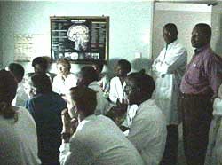 |
| Morning conference in radiology with the clinicians, where all cases of interest are presented and the clinical treatment discussed. |
The daily work begins at 7:30 when the various clinical teams (i.e. internal medicine, orthopedics, pediatrics, etc.) arrive in the radiology department, and cases from the previous day are discussed at length. Interns and residents present the cases, and the radiographic findings from the various studies are correlated with the patient's clinical information and treatment regimen.
Nearly all teams have some temporary members from the international community. Those countries represented during the month of October included Germany, Holland, England, Ireland, Norway, and the U.S. Case after case of pathology seldom or never seen in the U.S. are discussed and the clinical treatment plan is formulated by the teams after the cases are reviewed.
The most heavily utilized modalities in the department are ultrasound and plain-film radiography. Although helical CT is available, ultrasound is the mainstay of cross-sectional imaging. The advantages of lower cost, portability, and reliability give ultrasound the advantage in a country where radiology equipment service is extremely difficult to obtain and where the cost of a CT scan of the head is approximately 50% the average annual income of a Tanzanian worker.
As mentioned previously, the ultrasound machines used in the department span the quality spectrum, and every available unit, regardless of capability, is utilized. Some of the older units include an ATL/ADR 4000 S/LC, a Medison scanner, and a Philips SDR 3250. A recently acquired Hewlett-Packard Sonos 100CF is the only unit with color Doppler capability and is used primarily by Dr. Diefenthal, who usually performs the five or more daily echocardiograms himself. He acts as the technologist, the clinician, and the radiologist by interpreting the electrocardiogram, performing ascultation, preparing the patient, acquiring the images, and interpreting the exam with the ease of a very experienced echocardiographer.
Over the course of my short stay, complex and interesting cases were abundant, including congenital heart disease (i.e. tricuspid atresia, Ebsteins Anomaly, and many septal defects), severe rheumatic heart disease, and a chronic tuberculous pericardial effusion that produced 7.5 liters (yes, 7.5 liters) of fluid when aspirated. Valvular dysfunction was nearly ubiquitous, and wall-motion abnormalities were often due to cardiomyopathies as well as coronary disease.
In addition to the traditional ultrasound exams of the abdomen, retroperitoneum and pelvis, non-cardiac ultrasound involved every conceivable body part including musculoskeletal exams, transcranial B-mode, and opthalmic ultrasound. Frequently, the number of non-cardiac exams exceeded 40 per day, with an incredibly high percentage of positive cases. Case after case of HIV abdominal manifestations, hepatic abscesses, and large solid organ malignancies were scanned, along with cases the staff found more interesting including an intracranial cystic mass (thought due to Echinococcus) and unusual presentations of bladder shistosomiasis.
Many of the ultrasonographic examinations were non-standard by U.S. criteria. For example, some of the referring pediatricians did not hesitate to order transcranial ultrasounds on children older than five years because of the high prevalence of intracranial masses so large that the overlying calvaria was thin enough to scan through using almost any available probe. Ultrasounds were also requested to evaluate colonic masses, knees, and long bones (looking for subperiosteal effusions indicating osteomyelitis).
In addition to being heavily utilized, ultrasound was readily accepted by the Tanzanian patients referred to KCMC. There are no ultrasound technologists in the department and all of the exams are performed by the residents, assistant medical officers, and physicians. Comparatively, the doctor-patient communication seems superior to the traditional practice in the U.S., and physical exam findings and clinical histories are more easily correlated to the imaging findings (which is fortunate, since some findings need to be seen to be believed).
Another attraction to ultrasound for the patients is its affordability. The average price of an examination is 3,000 Tanzanian Shillings (equivalent to $3.80). This is within the range of what can be purchased by the average individual within the region, and is more than 10 times less than the price of a non-contrast CT scan of the head.
Apart from the quality of the scan and the price, the hospitalization experience can be fairly traumatic for the average Tanzanian patient due to the shortage or complete lack of items that facilitate patient comfort. Items such as sedatives and local anesthetics are rarely used, and patients are usually pleasantly surprised to find out that their ultrasound examination does not cause pain. The exam, therefore, is seen as a short but pleasant respite from the otherwise fairly grisly experience.
Plain-film radiography is, by developing country standards, excellent. Although the film-screen combinations are not ideally matched due to film availability constraints and the higher price of rare earth screens, the radiographs taken in the department are of good quality and are magnitudes better than the outside films brought by patients who have been seen at surrounding hospitals.
Every day there are about 60 radiographic exams, and almost every case demonstrates some type of pathology. Severe fractures are ubiquitous, and the morbidity associated with these fractures is substantial. The post-operative infection rate is high, and the metallic fixation devices very limited. Consequently, nearly all closed fractures (no matter how complex or in what location) are treated with closed reduction.
Many cases of fused joints and osteomyelitis due to trauma or surgery are seen daily. Patients commonly have draining sinuses from their infections and can have persistent drainage for years at a time. Many of these fractures, which would be treated by compression screws or plate fixation in the Unites States, are treated with traction and time, and a large percentage of these end with malunions or non-unions.
Cases seldom or never seen in the western world, such as rickets or congenital syphilis, are seen with regularity at the KCMC. Plain-film diagnoses of congenital heart disease is not unusual, and manifestations of untreated disease is routine. Due to the lack of appropriate chemotherapy, advanced appearances of various malignancies can be striking and unrecognizable to a radiologist accustomed to practicing in the U.S. The presence of cross-sectional imaging has proven to be very beneficial in evaluating some of the very complex disease that is commonly seen.
Computed tomography at the KCMC is an interesting mix of modern technology and Third World limitations. Although the unit (a Philips Tomoscan SR 4000) is modern and has helical capability, the cost to maintain the unit makes its operation an exercise in diminished return.
Contrast is very expensive by Tanzanian standards (equal to two weeks of a residents salary for 100cc) so enhanced abdominal scans are very rarely done. The workload is about 70% head CTs and, although many more studies are needed, most patients simply can't afford the cost of the exam. The result of these limitations is that only a fraction of the scanner's capability is used on the very small fraction of people that could benefit greatly from the information gathered from the exam.
A unique and very interesting component of the interaction with the patient involves "the morning march to x-ray." Although patient scheduling for all modalities has been attempted, the patients arrive en masse each morning to wait for their exam. Some of these patients will wait up to six hours before they are called but, for most of the patients, this waiting time also provides the social interaction that the Tanzanian people consider very important.
Most patients are ambulatory, and many of the visibly debilitated patients will even refuse wheelchair transport in order to show that they are strong enough to walk. They occupy the waiting room and the outside hallway benches, where they are called in turn for their examination. This approach continues until mid-afternoon, when all of the patients have been seen. Add-on studies are taken during the day, and after-hours requests are triaged by the resident on call.
The practice environment
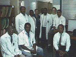 |
| The department of radiology at the Kilimanjaro Christian Medical Center. From left to right the individuals include: John Kachenesa, AMO; Cuthbert Kiluwa, AMO; E. Minja, M.D.; Helmut Diefenthal, M.D.; Rha Josiah, M.D.; Chrispin C. Moshi, M.D.; Suzi Muir, M.D. (visiting resident from UCLA); Ahmed M. Jusabani, M.D., and Alpha P. S. Lyimo, M.D.; not pictured (Ignace Masawe, AMO) |
As good as the past decade has been for Dr. Diefenthal and company, other hospital-based departments, such as laboratory and pharmacy, have not matched the recent improvements in radiology at the KCMC. The lack of adequate support services has been a substantial handicap to providing timely and accurate patient care. Basic antibiotics, such as penicillin and sulfa derivatives, are available, as are antimalarial medications and first-line medications for tuberculosis.
More recently developed and wide-spectrum antibiotics are not available, and treatment for some endemic conditions, such as schistosomiasis, is commonly in short supply. Medical therapy for HIV and chemotherapy for malignancies is not available, and the prolonged administration of any medication can be prohibitively expensive.
The laboratory provides only the basic blood profiles, and extended chemistries, including other slightly more complex assays, are not available. The microbial cultures done in the laboratory are poor at best and a comment by one of the internal medicine physicians, "I've been here four years and I've never seen anything grow from anything ... except for contaminants," best summarizes the situation.
The blood bank is as equally ill-equipped, and more than once responded to physician requests for blood by stating, "If you want your patient to have blood, you'll have to provide it yourself." Many of the support services taken for granted in the U.S. are either unavailable or are of marginal quality. The feeble state of these and other services (i.e. medical equipment repair, hospital engineering, janitorial support, etc.) and its unfavorable effect on the radiology department emphasizes our reliance on a large number of hospital services to provide high-quality patient care.
There are other difficulties with the local practice environment. While visiting radiologists are allowed to teach residents and to provide assistance with interpretations, they are not allowed to participate directly in the daily work environment and cannot generate written reports. The application for a work permit and Tanzanian license can be extremely difficult, and the initial request for the permit must come from a Tanzanian institution. This creates an unusual situation, in which visiting staff is not allowed to provide substantive services in an environment where this type of expertise is desperately needed.
With Dr. Diefenthal performing his own ultrasound exams and Dr. Lyimo's limited experience with ultrasound, there are cases performed by residents that simply cannot be checked due to time constraints and the lack of an adequate storage medium documenting the study. Despite the limitations, the benefit of a visiting radiologist to the department and to the patients is immense. The visitors are relied upon by the radiology residents and by the personnel from other departments for their diagnostic expertise and their imaging experience.
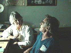 |
| Dr. Diefenthal consulting with visiting professor Dr. Genevieve Scarisbrick, a radiologist from Barrow-in-Furness, U.K. She is working at KCMC for six months for the purpose of providing humanitarian aid. |
The practice environment will be foreign, in both the literal and figurative senses, to a visiting radiologist accustomed to a Western style of practice. The working conditions are substantially more austere and the high prevalence of disease is accentuated by the advanced state of disease seen in a very high proportion of patients referred to the medical center.
As in many African countries, there is a high prevalence of HIV, and it has been estimated that 1 in 7 citizens have been infected with the virus. Although this rate is comparatively less than in some of the neighboring countries, the wards are replete with patients suffering from various opportunistic infections, and severe manifestations of the immunocompromised are common given the lack of effective medical therapy.
In addition to the limitations imposed by the paucity of medications or equipment, inertia within the practice environment is also a difficult barrier to surmount. Visitors are naturally looked upon with a certain amount of skepticism given their varied backgrounds and limited time commitments.
While most any practice could benefit from a fresh perspective, visitors to KCMC must consider the cultural and socioeconomic differences that create unique barriers to progress in this region. For example, the reluctance to discuss the HIV epidemic is one of the major reasons why the rate of infection is so high in many of the East African countries. Visitors from Western countries, however, must keep in mind that the societal acceptance of the disease is very different, and when the local physicians will not even discuss HIV infection openly there is no sense in considering the immediate introduction of a condom distribution program.
In a country with only nine radiologists there is also some reluctance within the establishment to accept outside influence. Given that supply is much less than the demand, the modern full-service practice espoused by Dr. Diefenthal has not been entirely popular with some of individuals content with a near monopoly on radiology services.
The benefits of this approach, however, are self-evident to the referring clinicians and patients, and this type of modern imaging practice provides makes a much greater impact than an analogous type of practice in a more developed country. The current dependence on this type of a system was dramatically illustrated by the ecstatic expressions on the faces of the clinicians and the extremely warm welcome from the support staff when they saw that Dr. Diefenthal had returned from his six-week sojourn in the U.S.
Summary
If much of Western medicine is evidence-based, most of African radiology is reality-based, with constant harsh reminders of the condition of medical and radiological services in the Third World. First World medicine simply does not work under Third World conditions for a number of reasons. Some of those barriers to improving radiology service include the lack of adequate equipment service and trained personnel, inability to generate adequate revenue to pay for equipment operation, and the lack of adequate support services such as pharmacy and laboratory.
In addition to the medical practice parameters, many things we do not even consider in the U.S. are significant problems in Third World countries (i.e., electricity, communication, political instability, etc.) The Diefenthals have managed, through teamwork and persistence, to bring East African radiology out of a primitive state and create an environment where high-quality imaging can be practiced in an austere environment.
Significant, but not insurmountable, barriers remain to realizing the full potential of the application of radiology in Tanzania. Critical to that success will be an increased willingness of volunteer radiologists to provide humanitarian aid and increased participation by the radiologic equipment corporations to donate and/or maintain the needed equipment.
If these factors can be combined with a long-term commitment by a radiologist willing and able to uphold the tradition of excellence established by Dr. Diefenthal, radiology will continue to flourish in East Africa for many years to come. In the meantime, a guaranteed once-in-a-lifetime experience is available for any radiologist that has the time and desire to visit this wonderful country.
Notes:
The Web site for the East African Medical Assistance Foundation can be found at www.eastafricafoundation.org.
Address of the EAST AFRICA MEDICAL ASSISTANCE FOUNDATION, c/o Dr. John Knoedler, president, 14 Island Road, North Oaks, MN 55127, USA. The foundation is registered with the IRS as a tax-exempt charitable organization.
The American Board of Radiology accepts three months at KCMC as part of its radiology residency requirements.
Kilimanjaro School of Radiology operates under the aegis of the Kilimanjaro Christian Medical College, which is one of the two colleges of the Tumaina University.
